©The Author(s) 2023.
World J Stem Cells. May 26, 2023; 15(5): 476-489
Published online May 26, 2023. doi: 10.4252/wjsc.v15.i5.476
Published online May 26, 2023. doi: 10.4252/wjsc.v15.i5.476
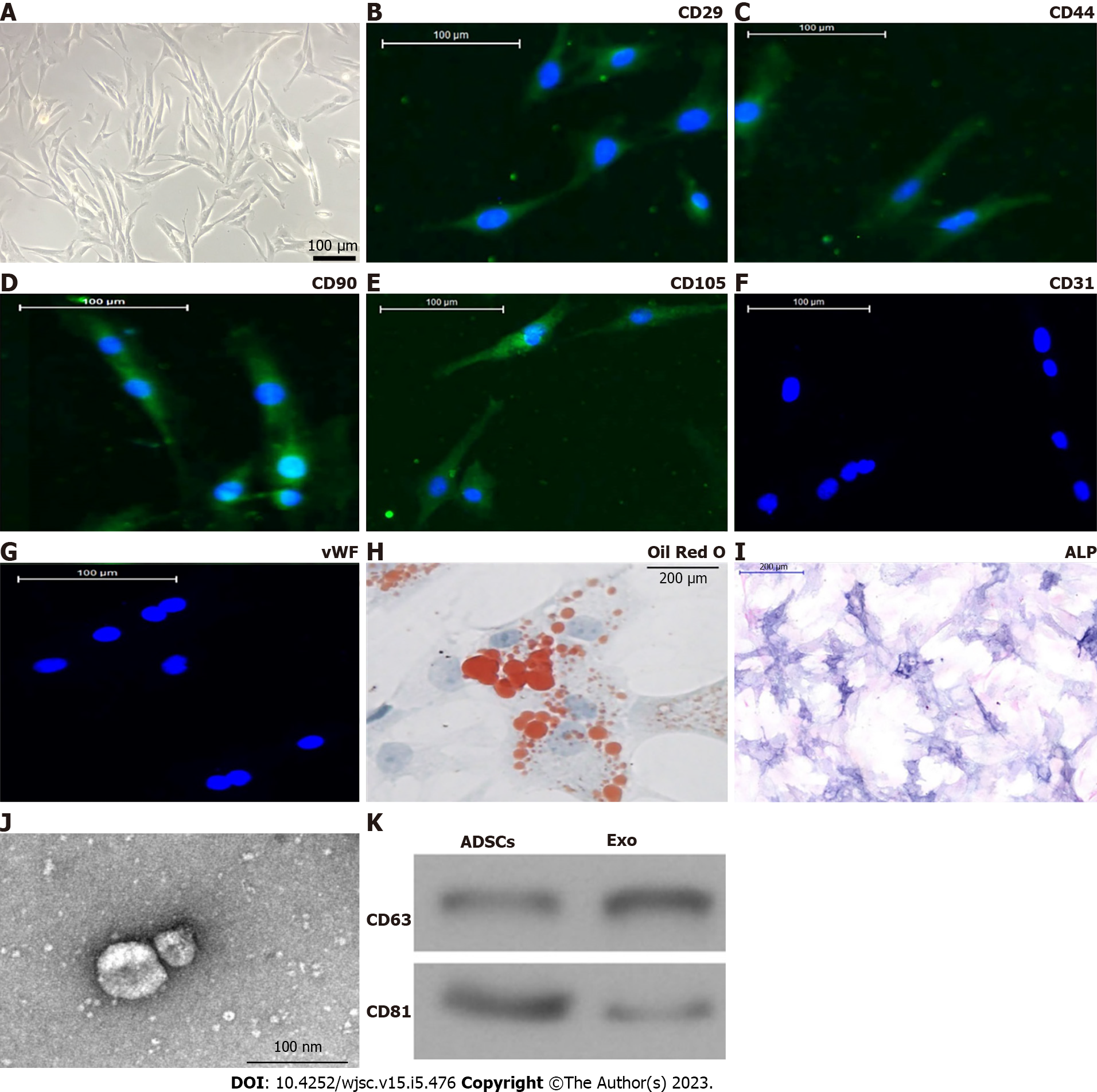
Figure 1 Characterization of adipose-derived mesenchymal stem cells and exosomes.
A: Adipose-derived mesenchymal stem cells (ADSCs) showed a typical cobblestone-like morphology. Scale bar: 100 μm; B–G: Immunofluorescence staining of cell surface markers. ADSCs exhibited positive expression of cluster of differentiation 90 (CD90), CD29, CD44, and CD105, but not von Willebrand factor or CD34. Scale bar: 100 μm; H and I: The differentiation potential of ADSCs assessed by Oil Red O (H) and alkaline phosphatase (I) staining. Scale bar: 200 μm; J: Transmission electron micrographs demonstrated ADSC exosome morphology. Scale bar: 100 nm; K: Western blotting detection of CD81 and CD63 expression in exosomes and ADSCs.
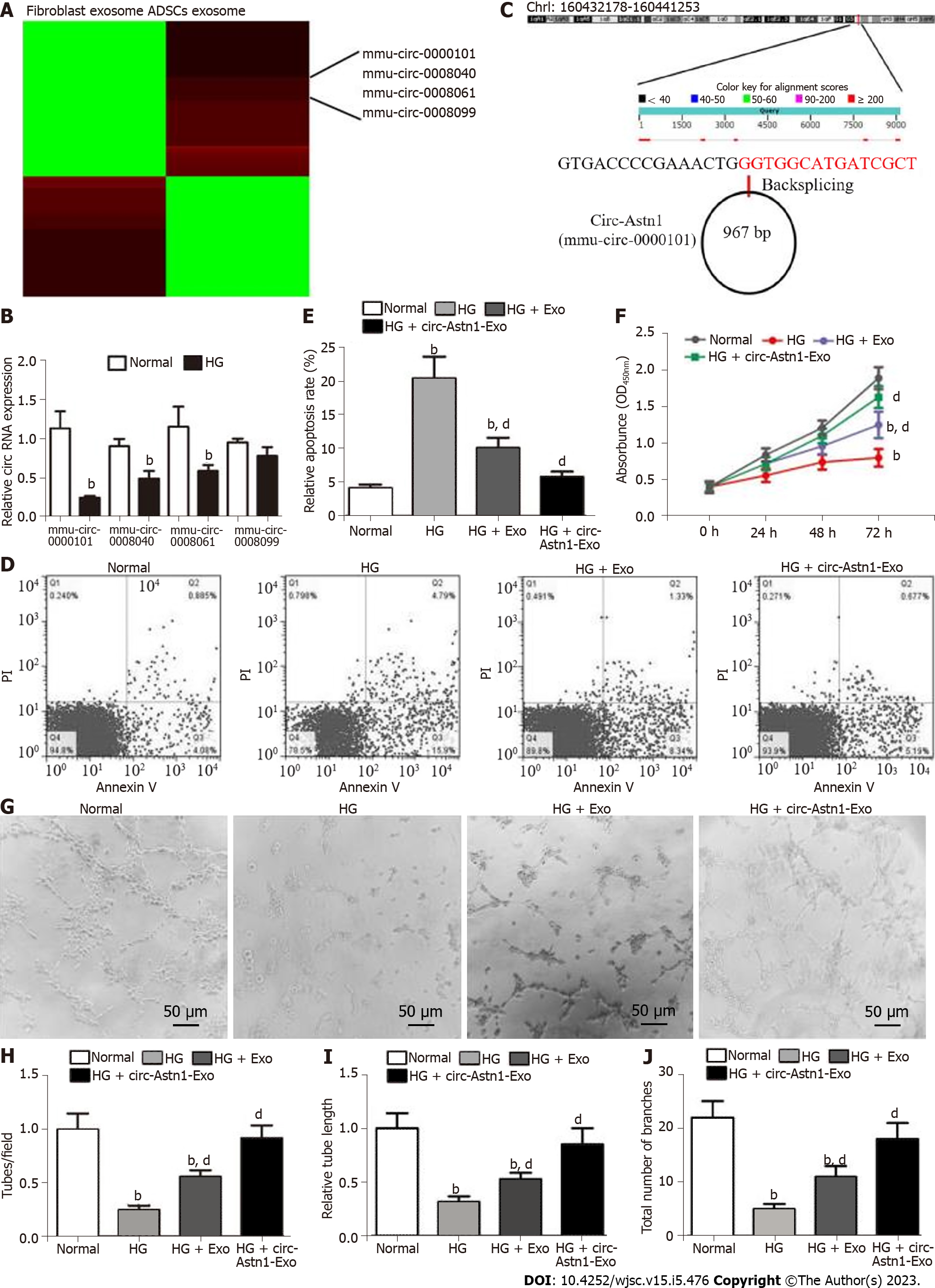
Figure 2 Exosomes derived from circular RNA astrotactin 1-modified adipose-derived mesenchymal stem cells function importantly in endothelial precursor cell function restoration by decreasing apoptosis under high glucose conditions.
A: Heat map regarding all differentially expressed circular RNAs (circRNAs) between adipose-derived mesenchymal stem cells (ADSCs) exosomes and fibroblast exosomes; B: Quantitative polymerase chain reaction giving mmu_circ_0000101 (circular RNA astrotactin 1), mmu_circ_0008040, mmu_circ_0008061, and mmu_circ_0008099 expression in endothelial precursor cells (EPCs) with or without high glucose (HG) treatment. Data are denoted by the mean ± SD; bP < 0.001 vs normal; C: The genomic loci of circ-Astn1; D and E: We pretreated EPCs with ADSC exosomes before treatment with exosomes for 1 d under HG conditions. Our team assayed EPC apoptosis via flow cytometry after annexin V-FITC staining. bP < 0.001 vs normal. dP < 0.001 vs HG; F: EPC proliferation under different treatments, determined by Cell Counting Kit-8 assay. bP < 0.001 vs normal. dP < 0.001 vs HG; G-J: Representative photomicrographs of tube-like structures. Scale bar: 50 μm. Technician-counted tube branch points (H), relative tube length (I) and the total number of branches were calculated. bP < 0.001 vs normal. dP < 0.001 vs HG. PI: Propidium iodide.
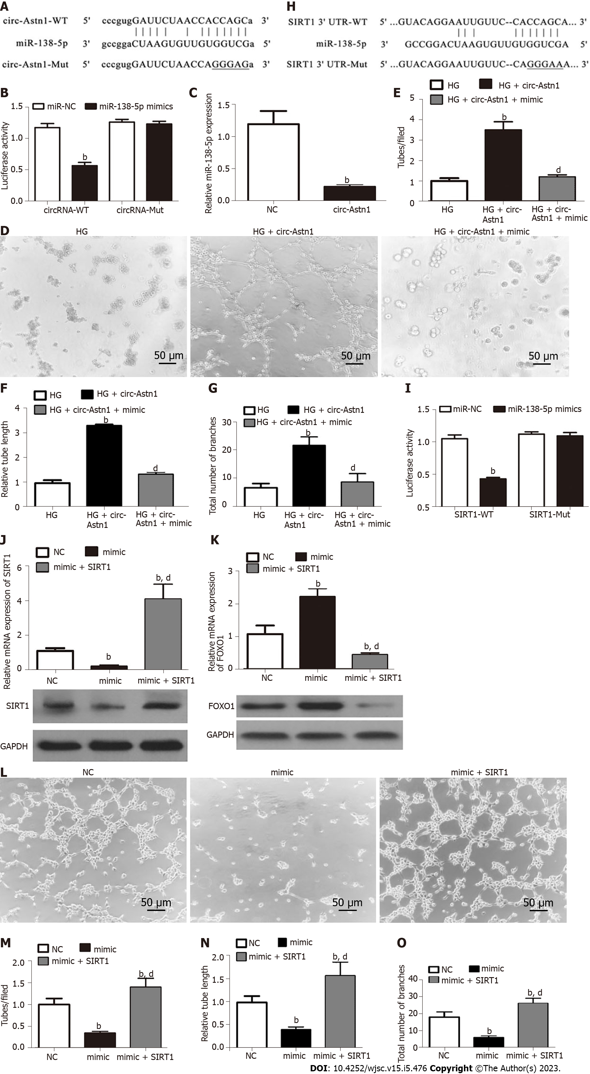
Figure 3 The circular RNA astrotactin 1-mediated mi-138-5p/SIRT1/forkhead box O1 signaling pathway plays an important protective role in endothelial precursor cells under high glucose conditions by promoting angiogenesis.
A and B: Luciferase expression levels in HEK293 cells transfected with cloned circular RNA astrotactin 1 (circ-Astn1) wild-type (WT) or mutant (MUT) vector and miR-138-5p mimics. Data are denoted by the mean ± SD. bP < 0.001; C: Quantitative polymerase chain reaction (qPCR) detection suggested that miR-138-5p expression was reduced after transfection with circ-Astn1-overexpressing vector in endothelial precursor cells (EPCs). Data are denoted as the mean ± SD. bP < 0.001 vs NC; D-G: Representative photomicrographs of tube-like structures of EPCs under high glucose (HG) conditions after transfection with negative control or circ-Astn1-overexpressing vector. bP < 0.001 vs HG. dP < 0.001 vs circ-Astn1; H and I: Luciferase expression level in HEK293 cells transfected with cloned SIRT1 WT- or MUT-3' UTR vector and miR-138-5p mimics. Data are denoted by the mean ± SD. bP < 0.001; J and K: qPCR and western blot analysis indicated that SIRT1 and forkhead box O1 expression were reduced after transfection with miR-138-5p overexpression vector in EPCs. Data are expressed as the mean ± SD. bP < 0.001 vs NC. dP < 0.001 vs miR-138-5p mimics; L-O: Representative photomicrographs of EPC tube-like structures under HG conditions after transfection with miR-138-5p mimics combined with or without SIRT1 overexpression vector. Data are denoted by the mean ± SD. bP < 0.001 vs NC. dP < 0.001 vs miR-138-5p mimics.
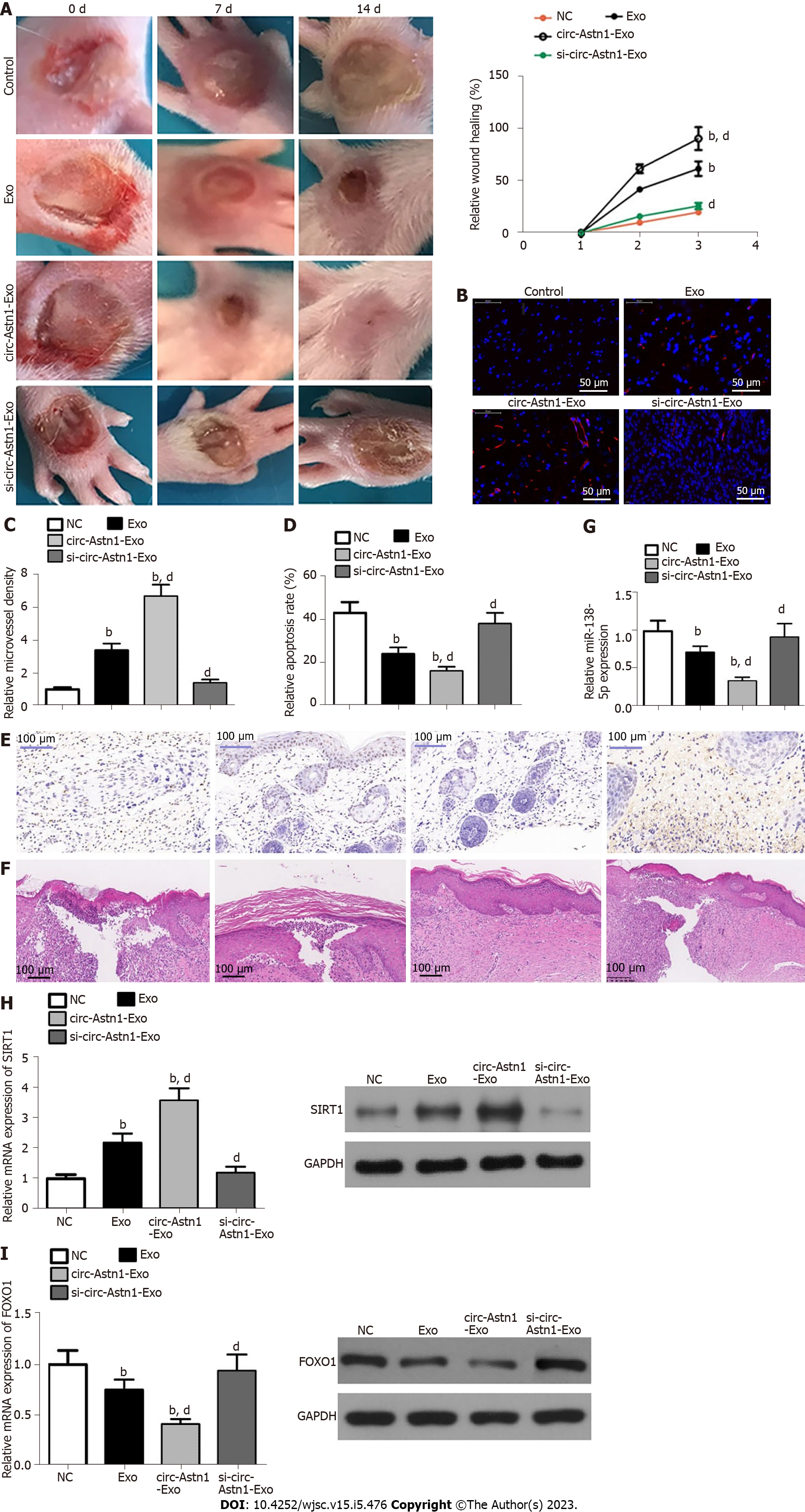
Figure 4 Exosomes from circular RNA astrotactin 1-modified adipose-derived mesenchymal stem cells have greater therapeutic effect in promoting wound healing in a diabetic mouse model.
A: Representative images of full-thickness skin defects after treatment with adipose-derived mesenchymal stem cell (ADSC) exosomes or circular RNA astrotactin 1-modified ADSC exosomes for 0, 1, and 2 wk after wounding; B and C: Microvascular formation evaluated by immunofluorescence staining with cluster of differentiation 31. bP < 0.001 vs control. dP < 0.001 vs exosomes; D and E: We assayed apoptosis level via terminal deoxynucleotidyl transferase dUTP nick end labeling staining. bP < 0.001 vs control. dP < 0.001 vs exosomes; F: Hematoxylin and eosin staining shows wound changes; G-I: Quantitative polymerase chain reaction and western blot analysis showing mi-138-5p (G), SIRT1 (H), and forkhead box O1 (I) expression. bP < 0.001 vs control. dP < 0.001 vs exosomes.
- Citation: Wang Z, Feng C, Liu H, Meng T, Huang WQ, Song KX, Wang YB. Exosomes from circ-Astn1-modified adipose-derived mesenchymal stem cells enhance wound healing through miR-138-5p/SIRT1/FOXO1 axis regulation. World J Stem Cells 2023; 15(5): 476-489
- URL: https://www.wjgnet.com/1948-0210/full/v15/i5/476.htm
- DOI: https://dx.doi.org/10.4252/wjsc.v15.i5.476


