©The Author(s) 2023.
World J Stem Cells. Mar 26, 2023; 15(3): 71-82
Published online Mar 26, 2023. doi: 10.4252/wjsc.v15.i3.71
Published online Mar 26, 2023. doi: 10.4252/wjsc.v15.i3.71
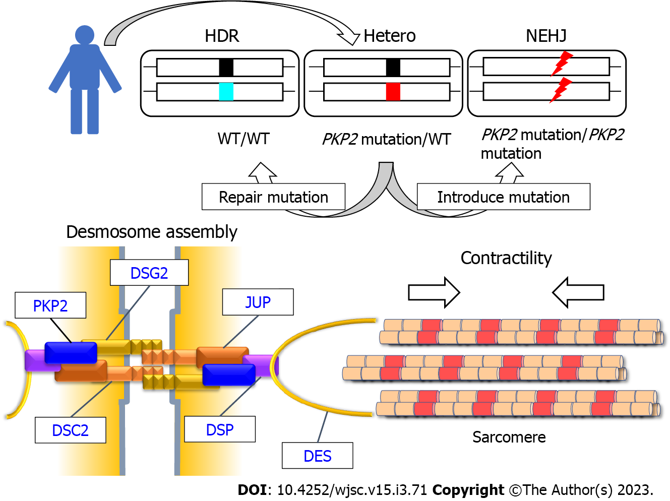
Figure 1 Modeling impaired desmosome assembly and reduced contractility using isogenic induced pluripotent stem cell-derived cardiomyocytes with the precisely adjusted dose of PKP2.
Heterozygous frameshift mutation in patient-derived induced pluripotent stem cells (iPSCs) was repaired through homology-directed repair. Homozygous frameshift mutations were introduced in PKP2 through non-homologous end joining in patient-derived iPSCs. The generated isogenic iPSC-derived cardiomyocytes with the precisely adjusted expression of PKP2 recapitulated impaired desmosome assembly and reduced contractility caused by PKP2 deficiency. Desmosomal cadherin proteins (DSG2 and DSC2) form homo-dimers and hetero-dimers. PKP2 is a scaffold protein for desmosomal cadherins, JUP, and DSP. Desmosomes are linked to sarcomere structure via the intermediate filament protein DES that targets both desmosome and Z disc structure. HDR: Homology-directed repair; NEHJ: Non-homologous end joining; Hetero: Heterozygous mutation.
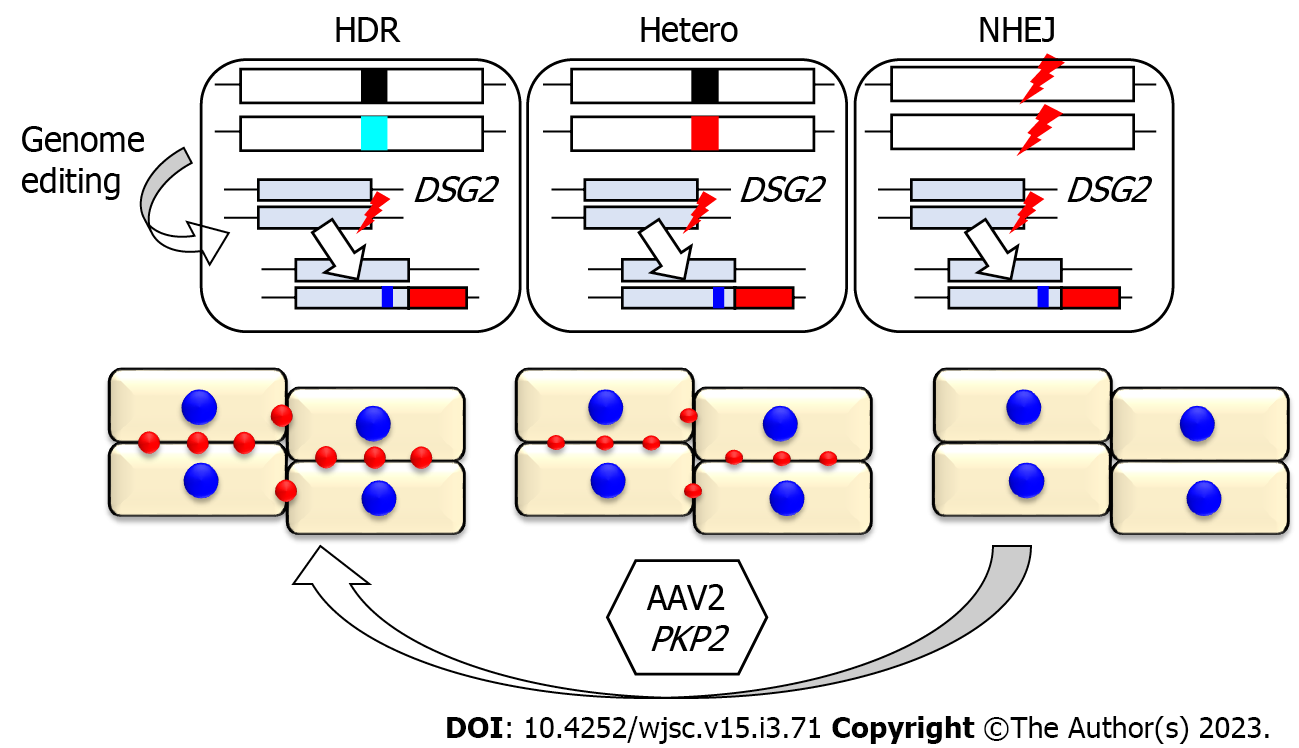
Figure 2 Allele-specific fluorescent labeling of DSG2 captures desmosome dynamics in isogenic induced pluripotent stem cell-derived cardiomyocytes.
To establish a model for desmosome imaging, the tdTomato fluorescent reporter was knocked-in at the 3′-terminus of DSG2 in the three established isogenic induced pluripotent stem cells (iPSCs) using genome editing. These isogenic iPSCs carried identical DSG2 alleles comprising intact and knocked-in alleles distinguished by a synonymous single nucleotide variant (indicated as blue line). These iPSC-derived cardiomyocytes enable desmosome imaging and capturing desmosome recovery after adeno-associated virus-mediated replacement of PKP2. HDR: Homology-directed repair; NEHJ: Non-homologous end joining; AAV: Adeno-associated virus; Hetero: Heterozygous mutation.
- Citation: Higo S. Disease modeling of desmosome-related cardiomyopathy using induced pluripotent stem cell-derived cardiomyocytes. World J Stem Cells 2023; 15(3): 71-82
- URL: https://www.wjgnet.com/1948-0210/full/v15/i3/71.htm
- DOI: https://dx.doi.org/10.4252/wjsc.v15.i3.71


