Copyright
©The Author(s) 2021.
World J Stem Cells. Aug 26, 2021; 13(8): 1005-1029
Published online Aug 26, 2021. doi: 10.4252/wjsc.v13.i8.1005
Published online Aug 26, 2021. doi: 10.4252/wjsc.v13.i8.1005
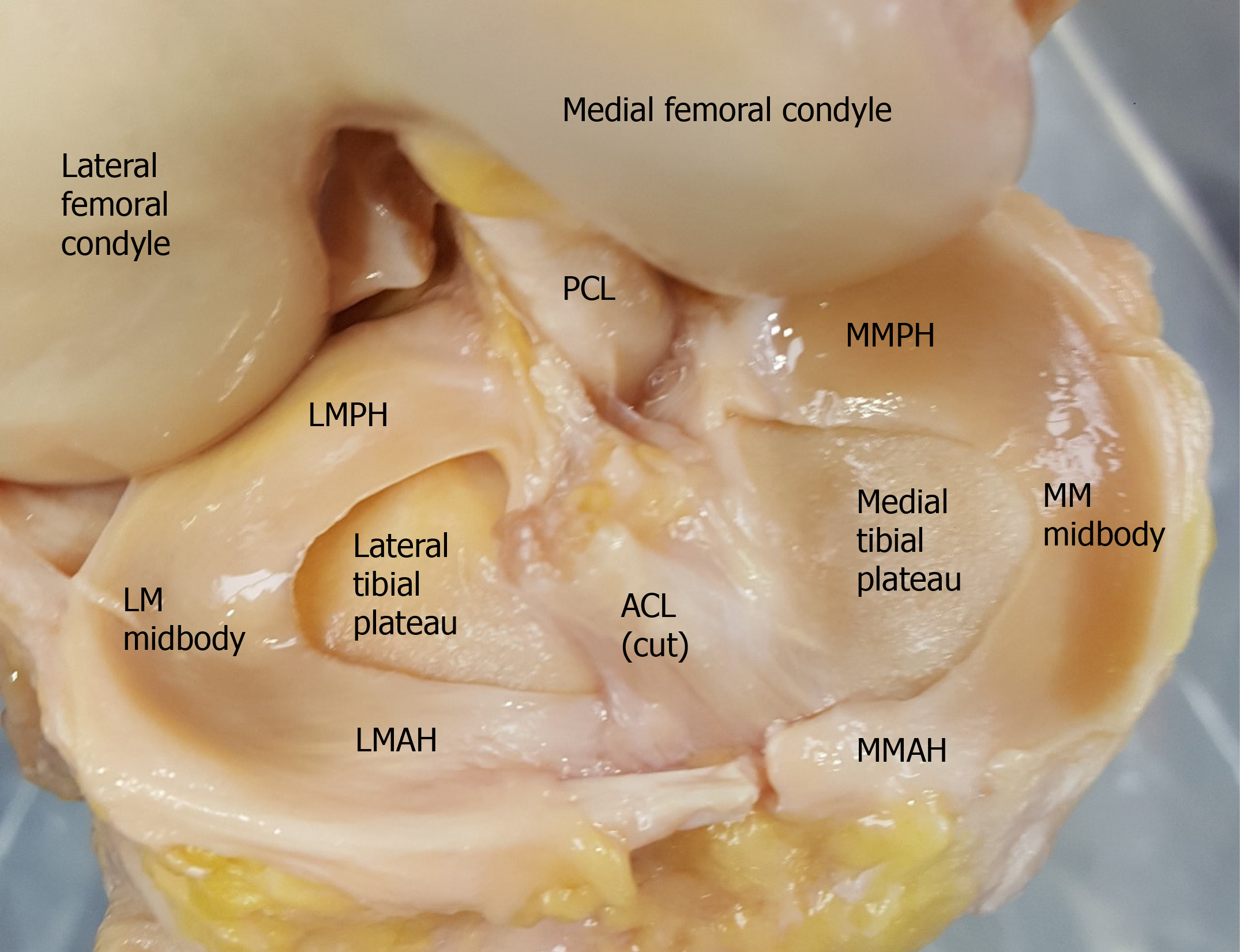
Figure 1 Anatomy of the meniscus in a cadaveric knee joint.
ACL: Anterior cruciate ligament; MM: Medial meniscus; MMAH: Medial meniscus anterior horn; MMPH: Medial meniscus posterior horn; LM: Lateral meniscus; LMAH: Lateral meniscus anterior horn; LMPH: Lateral meniscus posterior horn; PCL: Posterior cruciate ligament.
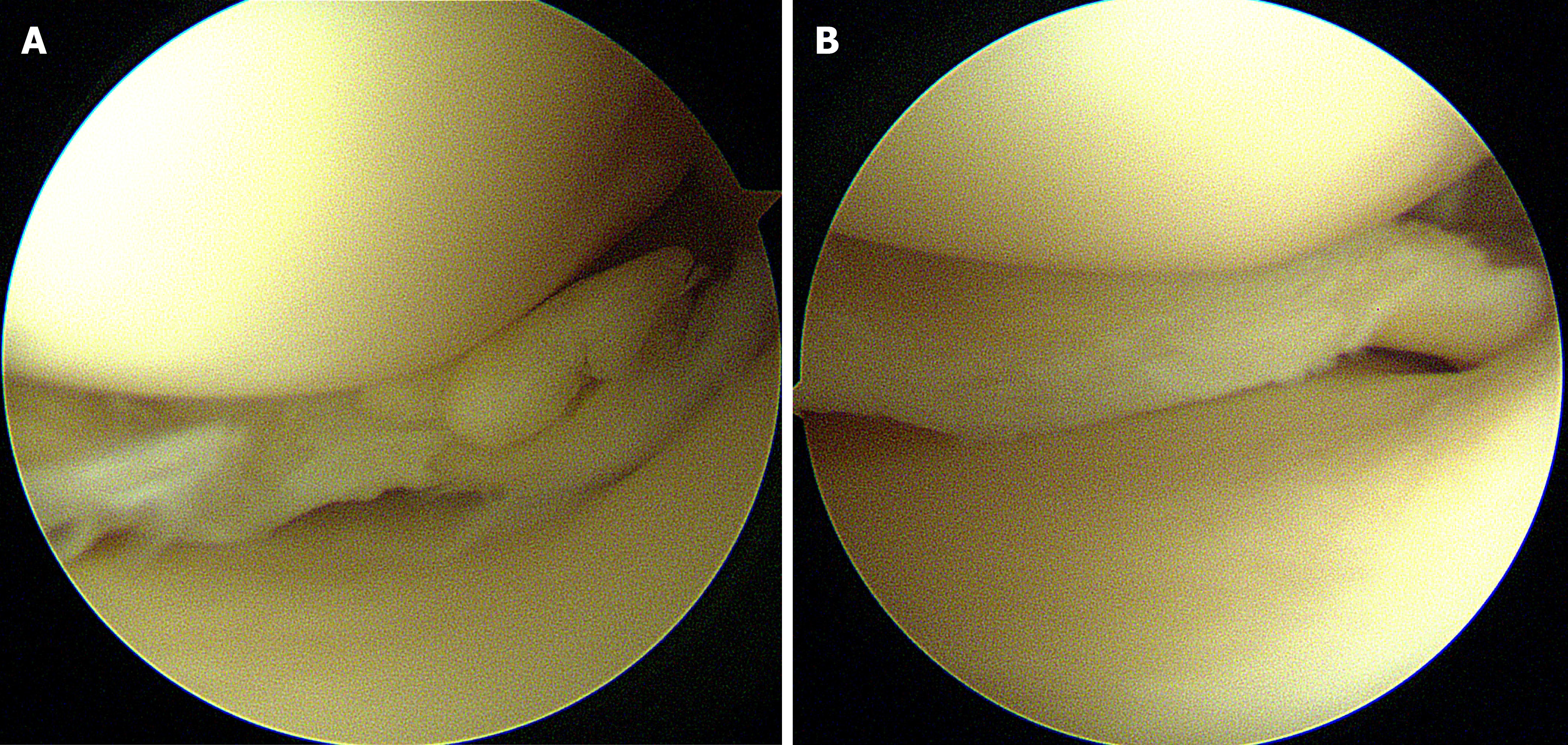
Figure 2 Arthroscopic images of partial meniscectomy.
A: Degenerative complex tear of the medial meniscus posterior horn; B: Remnant meniscus after partial meniscectomy.
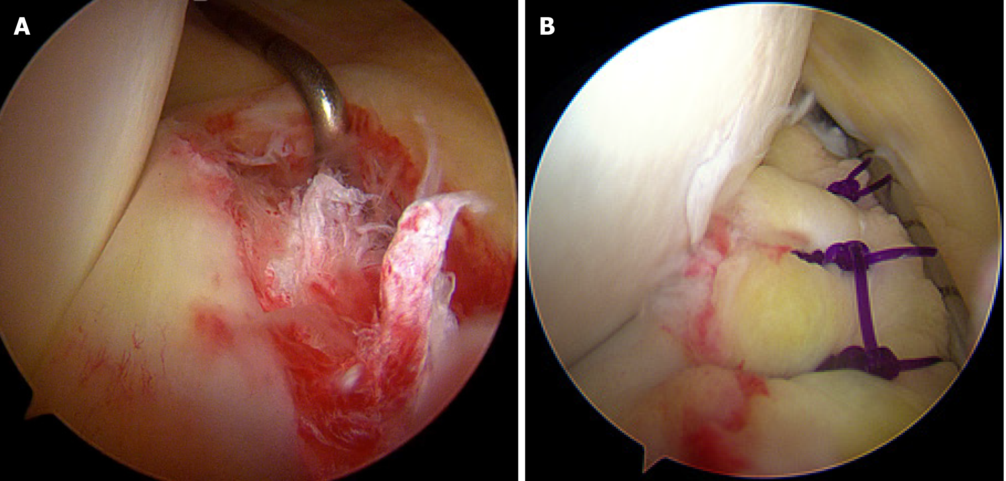
Figure 3 Arthroscopic images of meniscus repair.
A: Longitudinal tear in red-red zone of the medial meniscus posterior horn; B: Meniscus repair using all-inside suture technique.
- Citation: Rhim HC, Jeon OH, Han SB, Bae JH, Suh DW, Jang KM. Mesenchymal stem cells for enhancing biological healing after meniscal injuries. World J Stem Cells 2021; 13(8): 1005-1029
- URL: https://www.wjgnet.com/1948-0210/full/v13/i8/1005.htm
- DOI: https://dx.doi.org/10.4252/wjsc.v13.i8.1005


