Copyright
©The Author(s) 2021.
World J Stem Cells. Nov 26, 2021; 13(11): 1797-1812
Published online Nov 26, 2021. doi: 10.4252/wjsc.v13.i11.1797
Published online Nov 26, 2021. doi: 10.4252/wjsc.v13.i11.1797
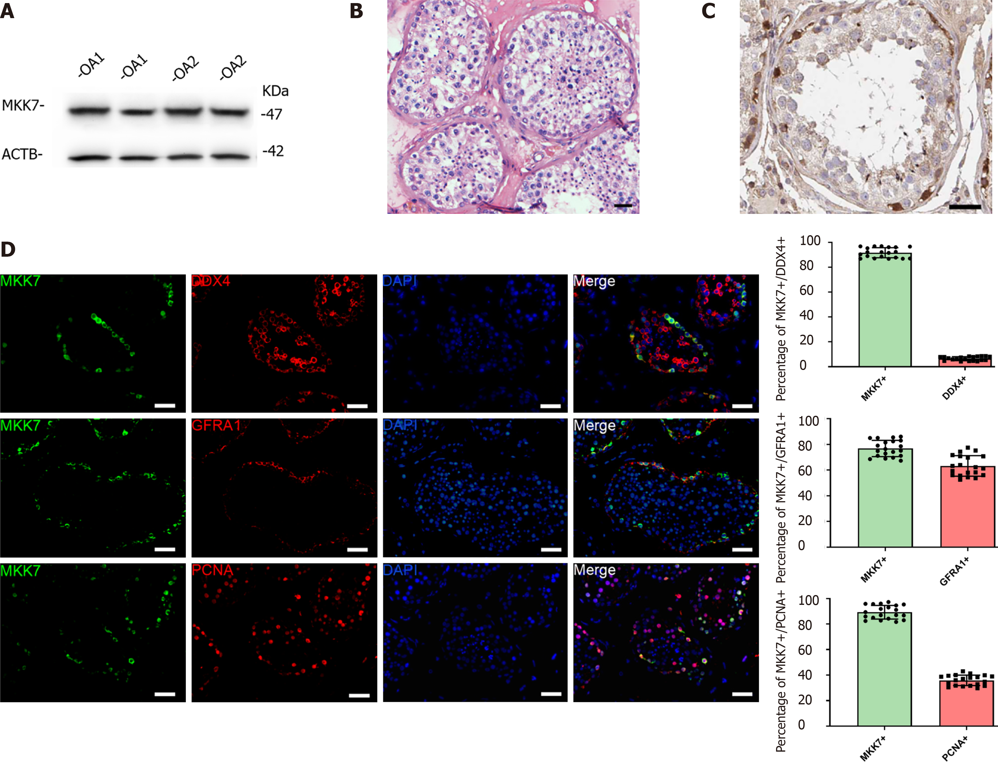
Figure 1 Mitogen-activated protein kinase kinase 7 was mainly expressed in human spermatogonial stem cells.
A: Mitogen-activated protein kinase kinase 7 (MKK7) was detected in samples from 2 patients with obstructive azoospermia (OA) using western blotting; B: Hematoxylin-eosin staining of testis with normal spermatogenesis; C: Immunohistochemistry revealing the cellular localization of MKK7 in normal testis; D: Double immunostaining showing the co-expression of MKK7 with DEAD-box helicase 4 (DDX4), glial cell line-derived neurotrophic factor family receptor alpha 1 (GFRA1) and proliferating cell nuclear antigen (PCNA) in normal human testis and the proportion of MKK7+ cells with DDX4, GFRA1 and PCNA expression; at least 20 seminiferous tubules were counted. Scale bars in B, C, and D: 50 μm. ACTB: Beta actin.
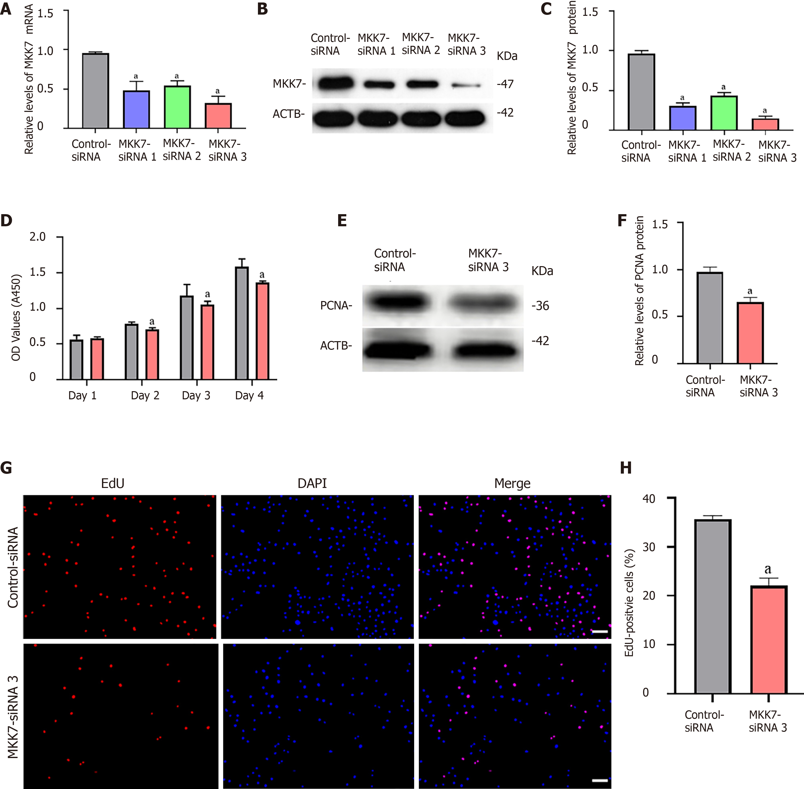
Figure 2 The influence of mitogen-activated protein kinase kinase 7 knockdown on human spermatogonial stem cell proliferation.
A: Quantitative real time PCR revealed mitogen-activated protein kinase kinase 7 (MKK7) mRNA level changes in a human spermatogonial stem cell (SSC) line after treatment with MKK7-small interfering RNA (siRNA) 1-, 2- and 3; B and C: Western blotting showed MKK7 protein level alterations in the human SSC line after the transfection of MKK7-siRNA 1-, 2- and 3. Beta actin (ACTB) was used as the loading control for the total protein; D: Cell counting kit (CCK)-8 assay illustrated the proliferation of human SSCs transfected with the Control-siRNA and MKK7-siRNA 3; E and F: The relative levels of proliferating cell nuclear antigen (PCNA) protein in human SSCs after transfection with the Control-siRNA and MKK7-siRNA 3; G and H: The percentages of EdU-positive cells in human SSCs transfected with the Control-siRNA and MKK7-siRNA 3. Scale bars: G, 50 μm. aP < 0.05 denotes a significant difference between the MKK7-siRNA 3 and Control-siRNA groups. DAPI: 4,6-diamidino-2-phenylindole.
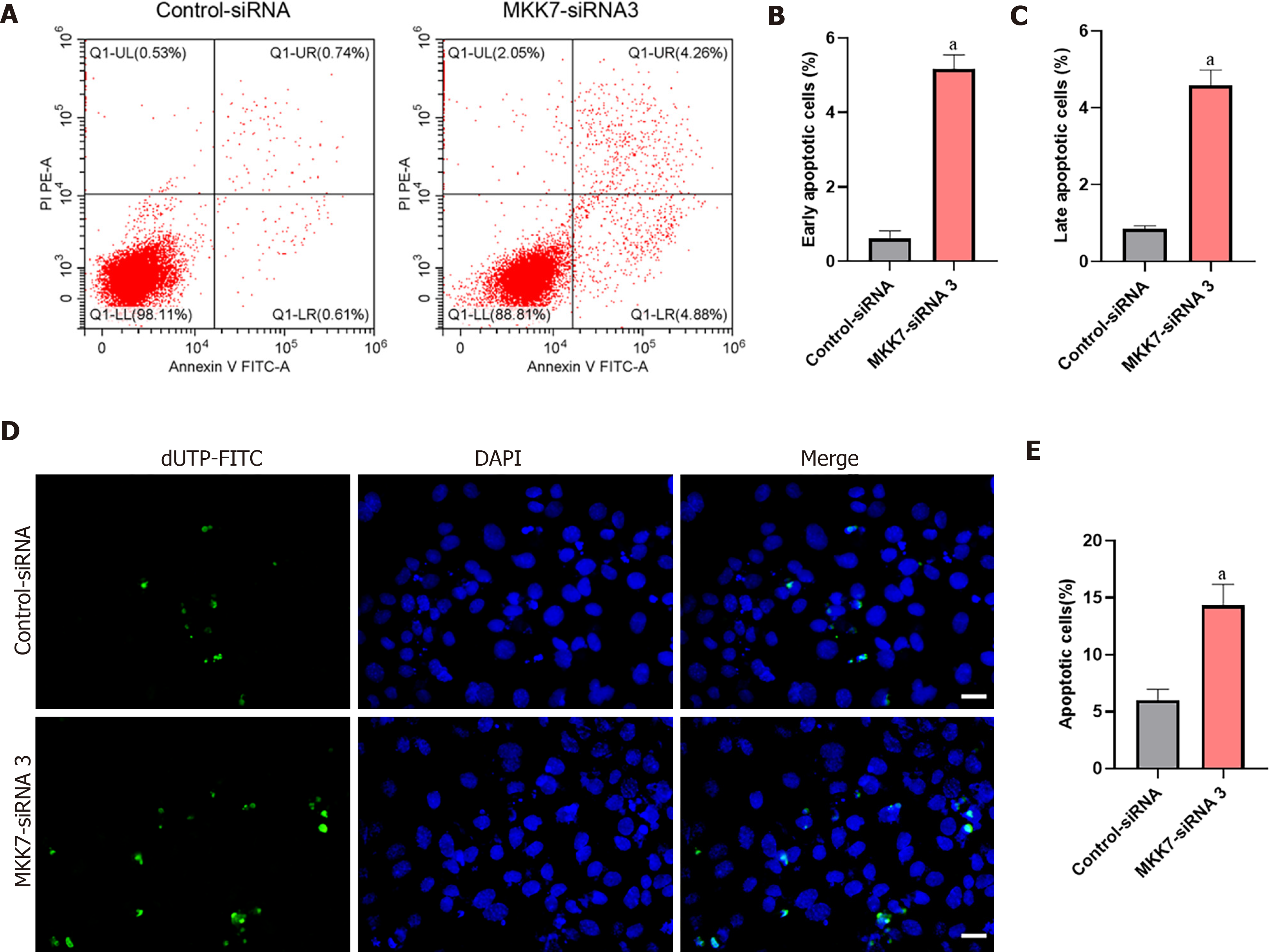
Figure 3 The influence of mitogen-activated protein kinase kinase 7 knockdown on the apoptosis of human spermatogonial stem cells.
A-C: Flow cytometry and fluorescein isothiocyanate (FITC) Annexin V analysis of the percentages of early (A and B) and late (A and C) apoptosis cells in human spermatogonial stem cells (SSCs) transfected with the Control-small interfering RNA (siRNA) and mitogen-activated protein kinase kinase 7 (MKK7)siRNA 3; D and E: Terminal deoxynucleotidyl transferase nick-end-labeling (TUNEL) assays of the percentages of TUNEL+ cells in human spermatogonial stem cells transfected with the Control-siRNA and MKK7-siRNA 3. Scale bars in D: 50 μm. aP < 0.05 denotes a significant difference between the MKK7-siRNA 3 and Control-siRNA groups. dUTP: Deoxyuridine 5’-triphosphate; DAPI: 4,6-diamidino-2-phenylindole.
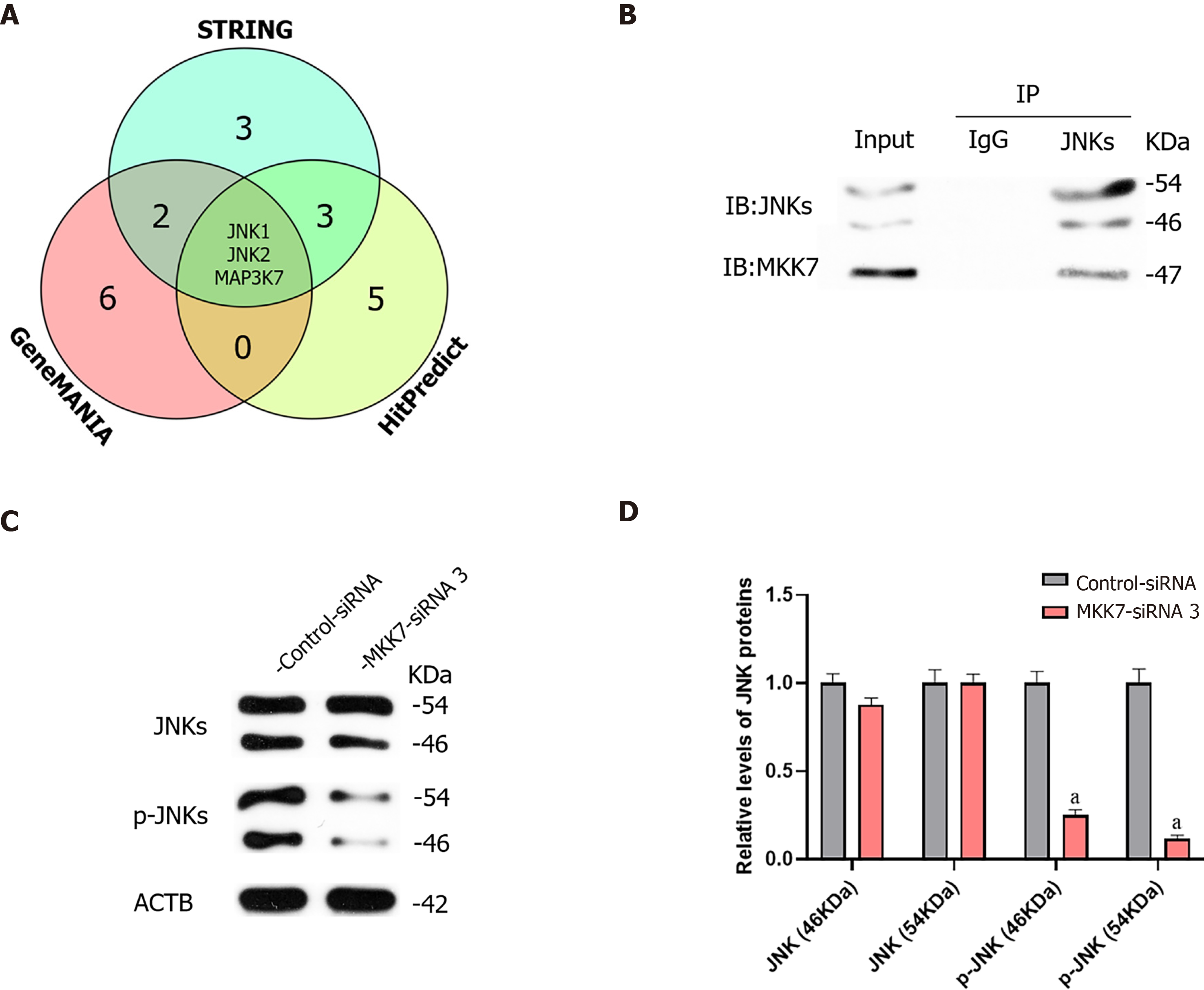
Figure 4 Mitogen-activated protein kinase kinase 7 affected the phosphorylation of c-Jun N-terminal kinases.
A: Three databases predicted that mitogen-activated protein kinase kinase 7 (MKK7) interacts directly with c-Jun N-terminal kinase 1 (JNK1), JNK2 and mitogen-activated protein kinase kinase kinase 7 (MAPK3K7); B: Protein co-immunoprecipitation (IP) results indicated that MKK7 interacts directly with JNKs; C and D: Western blotting results showed that MKK7 knockdown did not affect the overall expression level of JNKs; the levels of phosphorylated (p-)JNKs decreased significantly. aP < 0.05 denotes a significant difference between the MKK7-small interfering (si)RNA 3 and Control-siRNA groups. IB: Immunoblot; IgG: Immunoglobulin G; ACTB: Beta actin.
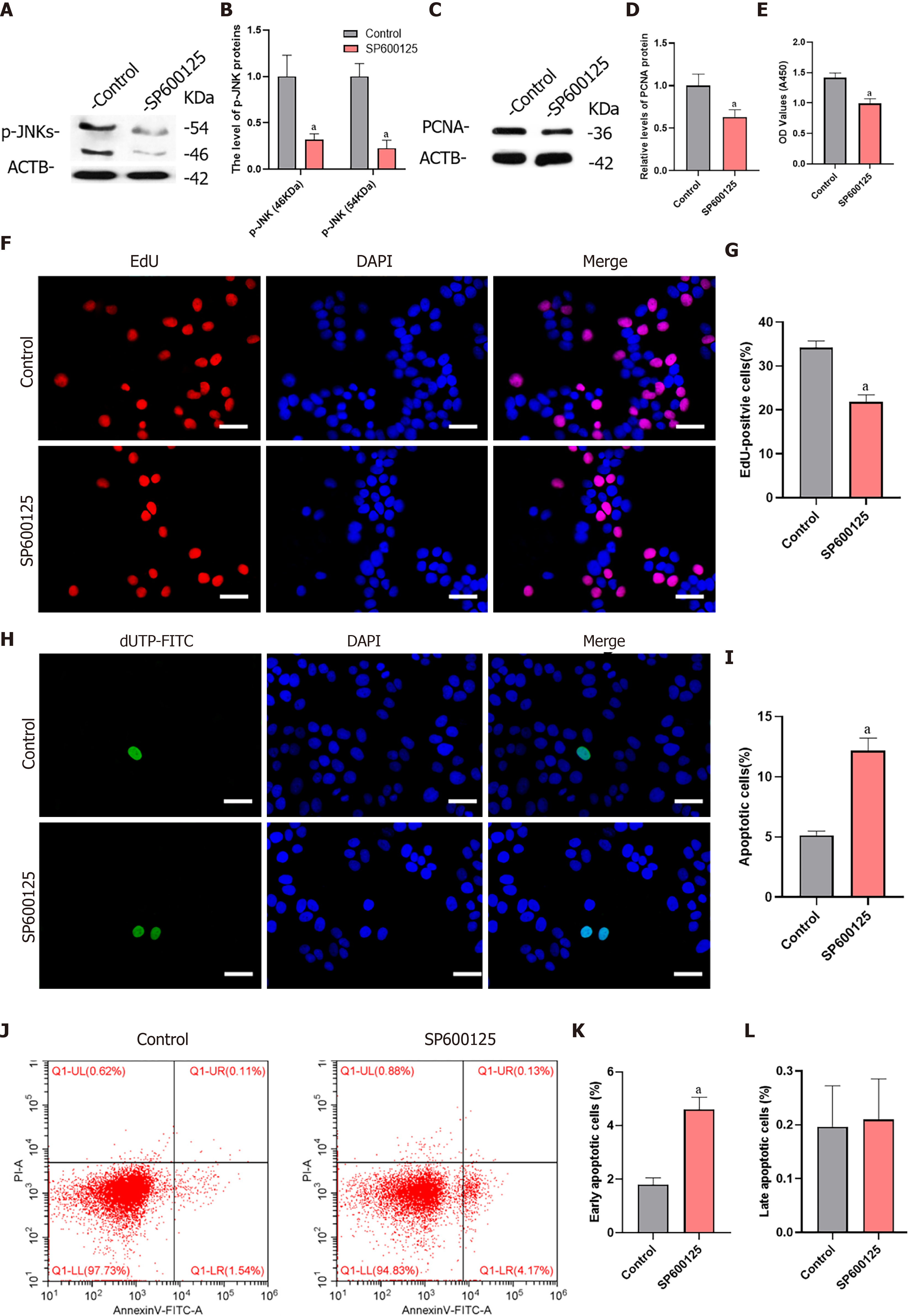
Figure 5 Inhibition of c-Jun N-terminal kinase 1 phosphorylation inhibited proliferation and promoted apoptosis of human spermatogonial stem cells.
A and B: Western blotting results showed that the phosphorylation of c-Jun N-terminal kinases was significantly inhibited by the c-Jun N-terminal kinase phosphorylation inhibitor, SP600125; C and D: The levels of proliferating cell nuclear antigen (PCNA) protein in human spermatogonial stem cells (SSCs) decreased significantly after treatment with SP600125; E: Cell counting kit (CCK)-8 results showed that the proliferation of SSCs was downregulated; F and G: EdU assays of DNA synthesis in human SSCs treated with SP600125; H and I: Terminal deoxynucleotidyl transferase nick-end-labeling (TUNEL) assay of the percentage of TUNEL+ cells in the human SSC line after SP600125 treatment; J-L: Flow cytometry and fluorescein isothiocyanate (FITC)/Annexin V analysis of the proportions of early (J and K) and late (J and L) apoptosis in the human SSC line treated with SP600125. Scale bars in F and H: 50 μm. aP < 0.05 denotes a significant difference between the Control and SP600125 treatment groups. p-JNK: Phosphorylated c-Jun N-terminal kinase; ACTB: Beta actin; DAPI: 4,6-diamidino-2-phenylindole.
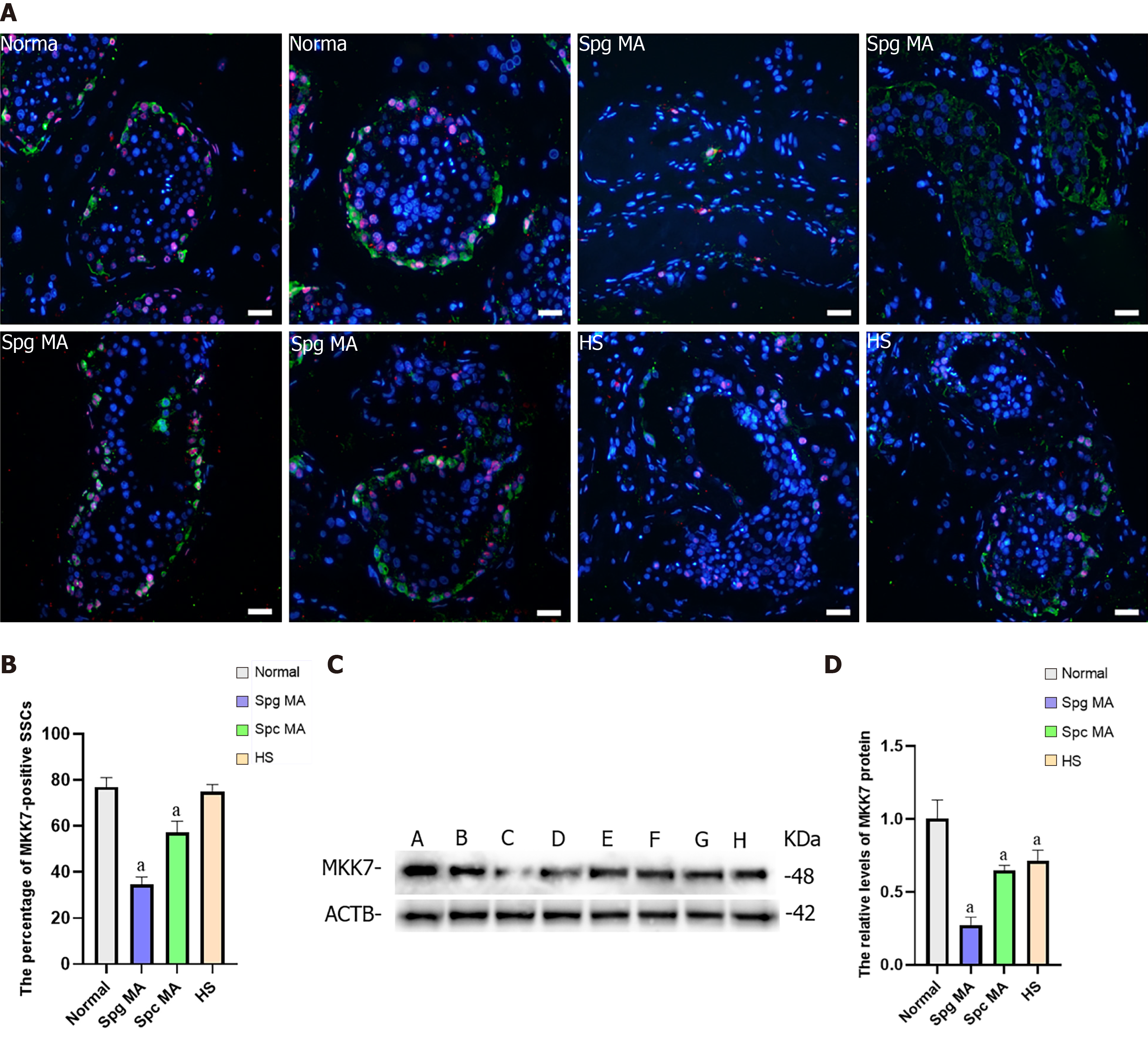
Figure 6 The expression of mitogen-activated protein kinase kinase 7 in the testis of obstructive azoospermia patients and non-obstructive azoospermia patients.
A and B: The percentages of ubiquitin C-terminal hydrolase L1 (UCHL1) (green)-positive spermatogonial stem cells (SSCs) with mitogen-activated protein kinase kinase 7 (MKK7) (red) expression between obstructive azoospermia (OA) and various kinds of non-obstructive azoospermia (NOA) patients; C and D: Western blotting compared the relative levels of MKK7 protein between OA and NOA patients. Notes in (C): sample A and B were individuals with OA with normal spermatogenesis; sample C and D were NOA patients with spermatogonia maturation arrest; sample E and F were NOA patients with spermatocyte maturation arrest; sample G and H were NOA patients with hypospermatogenesis. Scale bars: C, 50 μm. aP < 0.05 indicated the significant differences between OA patients with normal spermatogenesis and NOA patients. Spg MA: Spermatogonia maturation arrest; Spc MA: Spermatocyte maturation arrest; HS: Hypospermatogenesis; ACTB: Beta actin.
- Citation: Huang ZH, Huang C, Ji XR, Zhou WJ, Luo XF, Liu Q, Tang YL, Gong F, Zhu WB. MKK7-mediated phosphorylation of JNKs regulates the proliferation and apoptosis of human spermatogonial stem cells. World J Stem Cells 2021; 13(11): 1797-1812
- URL: https://www.wjgnet.com/1948-0210/full/v13/i11/1797.htm
- DOI: https://dx.doi.org/10.4252/wjsc.v13.i11.1797


