©The Author(s) 2020.
World J Stem Cells. Jul 26, 2020; 12(7): 621-632
Published online Jul 26, 2020. doi: 10.4252/wjsc.v12.i7.621
Published online Jul 26, 2020. doi: 10.4252/wjsc.v12.i7.621
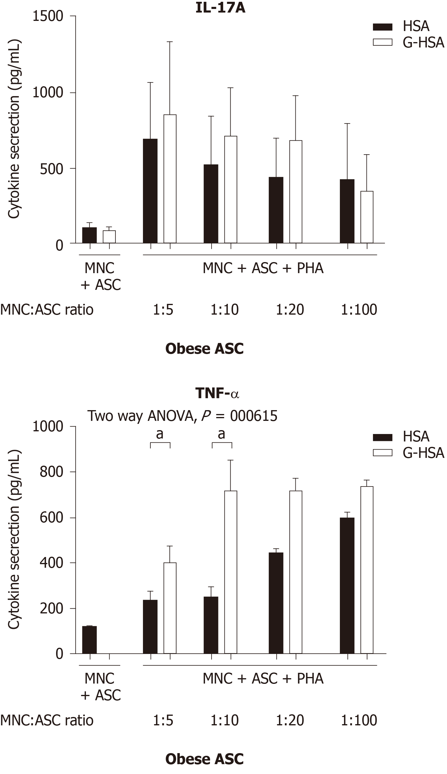
Figure 1 Glycated human serum albumin increases the levels of interleukin 17A promoted by obese adipose-derived stem cells, at suboptimal conditions.
Graded concentrations of obese adipose-derived stem cells (ASC) were co-cultured in the presence of mononuclear cells (MNC) at different ratios of 1:5, 1:10, 1:20, or 1:100, with 20000 ASC for 100000 MNC. Co-cultures were activated by phytohemagglutinin A (5 µg/mL) for 48 h in the presence of 1% human serum albumin or 1% glycated human serum albumin. ELISA were performed to measure interleukin-17A production and tumor necrosis factor alpha. Bars represent the mean ± SE of 4 independent experiments performed in triplicates. The P value shown in the figure corresponds to ANOVA multivariate analysis results, and aP < 0.05, as obtained by Bonferroni post-hoc tests. ASC: Adipose-derived stem cells; MNC: Mononuclear cells; PHA: Phytohemagglutinin A; HSA: Human serum albumin; G-HSA: Glycated human serum albumin; IL: Interleukin; TNF-α: Tumor necrosis factor alpha.
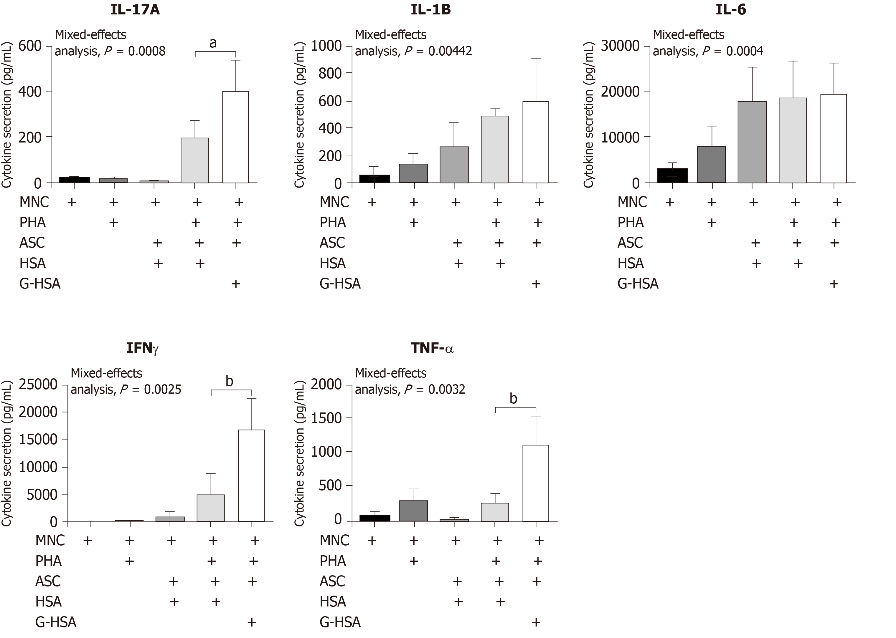
Figure 2 Lean adipose-derived stem cells increase the levels of interleukin 17A, tumor necrosis factor alpha, and interferon gamma secretion by mononuclear cells, in the presence of glycated human serum albumin.
Lean adipose-derived stem cells (ASC) were co-cultured with mononuclear cells (MNC) at the 1:5 ASC to MNC cell ratio, in the presence of 1% glycated human serum albumin or human serum albumin (HSA) for 48 h, phytohemagglutinin A (PHA) was added or not at 5 µg/mL. As control, MNC were cultured alone in the presence of PHA or not, and HSA. ELISA were performed to measure interleukin (IL)-17A, IL-1β, IL-6, interferon gamma, and tumor necrosis factor alpha production. Bars represent the mean ± SE of 5 independent experiments performed in triplicates. The P values shown in the figure correspond to ANOVA multivariate results and aP < 0.05, bP < 0.01, respectively as obtained by Bonferroni post-hoc tests. ASC: Adipose-derived stem cells; MNC: Mononuclear cells; G-HSA: Glycated human serum albumin; HSA: Human serum albumin; PHA: Phytohemagglutinin A; IL: Interleukin; IFNγ: Interferon gamma; TNFα: Tumor necrosis factor alpha.
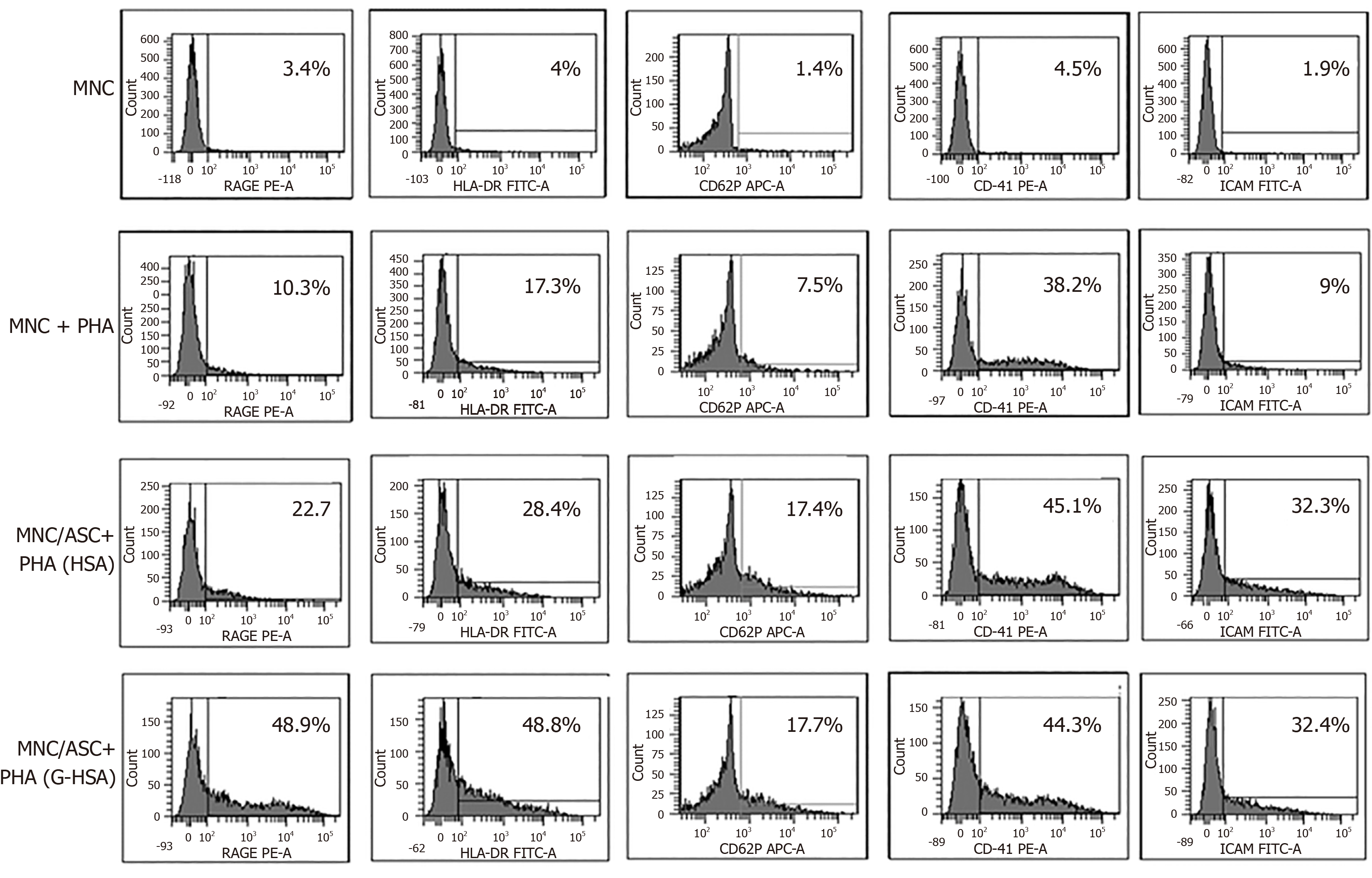
Figure 3 Glycated human serum albumin increases receptor for advanced glycated end products and human leukocyte antigen-DR expression in adipose-derived stem cell / mononuclear cell co-cultures.
Lean adipose-derived stem cells (ASC) were co-cultured with mononuclear cells (MNC) at the 1:5 ASC to MNC cell ratio, in the presence of 1% glycated human serum albumin or human serum albumin (HSA) for 48 h, phytohemagglutinin A (PHA) was added or not. As control MNC were cultured alone in the presence or not of PHA, and HSA. Human leukocyte antigen-DR, receptor for advanced glycated end products, cluster of differentiation (CD) 41, CD62P, and intercellular adhesion molecule 1 were stained with fluorescent-conjugated antibodies, and analyzed by cytofluorometry, using the DIVA software. This experiment is representative of two experiments performed, with two different ASC. ASC: Adipose-derived stem cells; MNC: Mononuclear cells; G-HSA Glycated human serum albumin; HSA: Human serum albumin; PHA: Phytohemagglutinin A; HLA: Human leukocyte antigen; RAGE: Receptor for advanced glycated end products; CD: Cluster of differentiation; ICAM-1: Intercellular adhesion molecule 1; FITC: Fluorescein isothiocyanate; PE: Phycoerythrin; APC: Allophycocyanin.
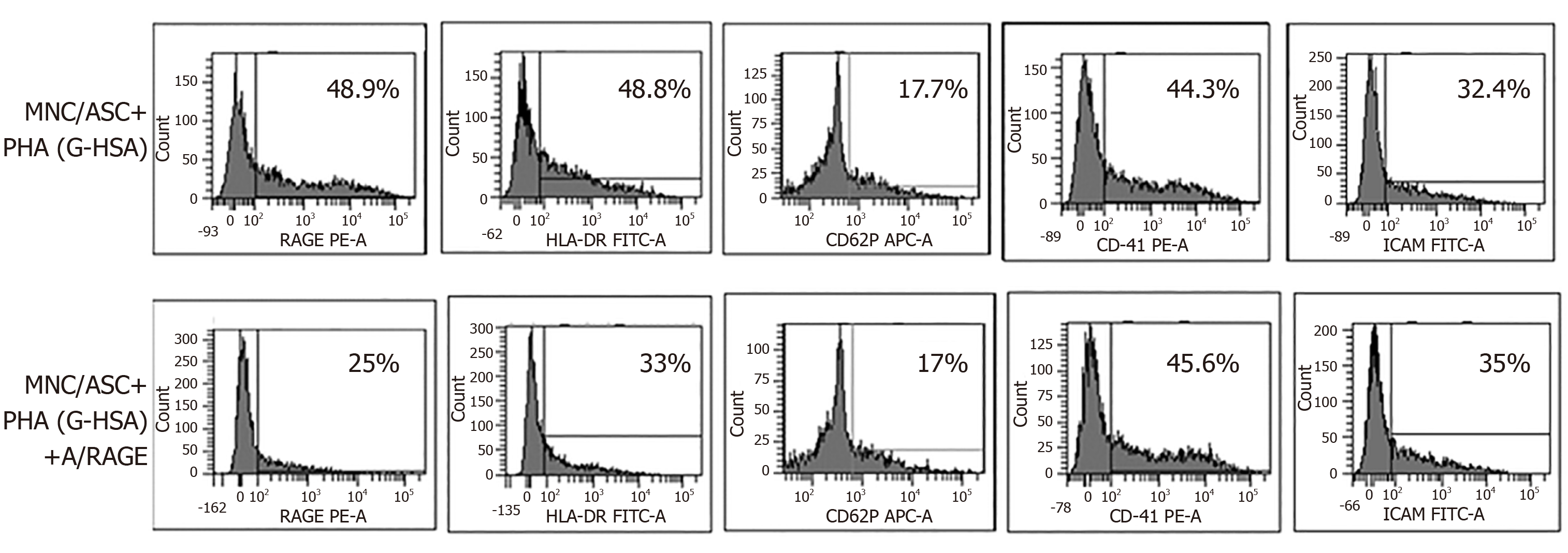
Figure 4 Anti- receptor for advanced glycated end products monoclonal antibody inhibits receptor for advanced glycated end products and human leukocyte antigen-DR expression.
Lean adipose-derived stem cells (ASC) were co-cultured with mononuclear cells (MNC) at the 1:5 ASC to MNC cell ratio, in the presence of 1% glycated human serum albumin for 48 h, phytohemagglutinin A was added at 5 µg/mL. Anti-receptor for advanced glycated end products (RAGE) monoclonal antibody was added at a concentration of 20 µg/mL. cluster of differentiation (CD) 41, CD62P, intercellular adhesion molecule 1, human leukocyte antigen-DR, and RAGE expression were stained with fluorescent-conjugated antibodies and analyzed by cytofluorometry, using the DIVA software. This experiment is representative of two experiments performed with two different ASC. ASC: Adipose-derived stem cells; MNC: Mononuclear cells; G-HSA: Glycated human serum albumin; PHA: Phytohemagglutinin A; RAGE: Receptor for advanced glycated end products; mAb: Monoclonal antibody; CD: Cluster of differentiation; ICAM-1: Intercellular adhesion molecule 1; HLA: Human leukocyte antigen; FITC: Fluorescein isothiocyanate; PE: Phycoerythrin; APC: Allophycocyanin.
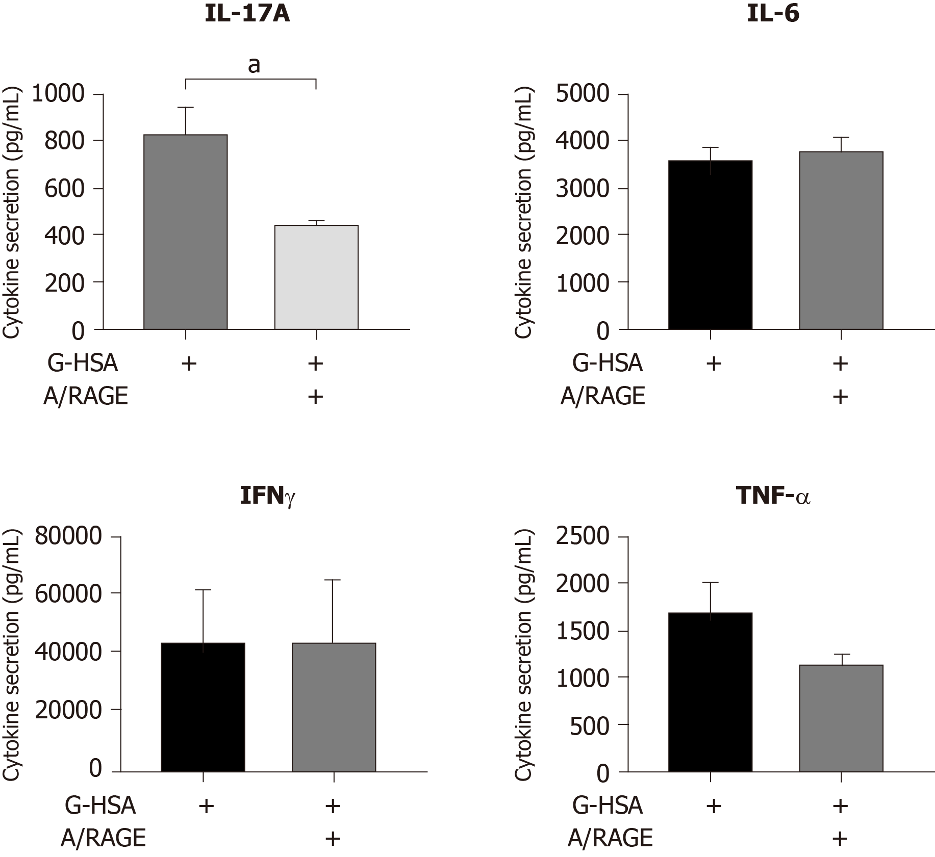
Figure 5 Inhibitory effects of anti-receptor for advanced glycated end products monoclonal antibody on interleukin 17A production.
Lean adipose-derived stem cells (ASC) were co-cultured with mononuclear cells (MNC) at the 1:5 ASC to MNC cell ratio, in the presence of 1% glycated human serum albumin for 48 h, phytohemagglutinin A was added. Anti-receptor for advanced glycated end products monoclonal antibody was added at a concentration of 20 µg/mL. ELISA were performed to measure interleukin (IL)-17A, interferon gamma, tumor necrosis factor alpha and IL-6 secretion. Bars represent the mean ± SE of 5 independent experiments performed in triplicates. aP < 0.05, as obtained by paired t tests. ASC: Adipose-derived stem cells; MNC: Mononuclear cells; G-HSA: Glycated human serum albumin; PHA: Phytohemagglutinin A; RAGE: Receptor for advanced glycated end products; mAb: Monoclonal antibody; IL: Interleukin; IFNγ: Interferon gamma; TNFα: Tumor necrosis factor alpha.
- Citation: Pestel J, Robert M, Corbin S, Vidal H, Eljaafari A. Involvement of glycated albumin in adipose-derived-stem cell-mediated interleukin 17 secreting T helper cell activation. World J Stem Cells 2020; 12(7): 621-632
- URL: https://www.wjgnet.com/1948-0210/full/v12/i7/621.htm
- DOI: https://dx.doi.org/10.4252/wjsc.v12.i7.621


