H Pylori Open Access
Copyright ©2005 Baishideng Publishing Group Inc. All rights reserved.
World J Gastroenterol. Jan 7, 2005; 11(1): 109-113
Published online Jan 7, 2005. doi: 10.3748/wjg.v11.i1.109
Effect of Helicobacter pylori VacA on gene expression of gastric cancer cells
Hong-Tao Wang, Zhen-Hong Li, Jian-Ping Yuan, Wei Zhao, Xiao-Dong Shi, Shan-Qing Tong, Xiao-Kui Guo, Department of Microbiology and Parasitology, Shanghai Second Medical University, Shanghai 20025, China
Author contributions: All authors contributed equally to the work.
Supported by the State Ministry of Education Research Foundation for Returned Overseas Chinese Scholars Abroad (2001) 498
Correspondence to: Xiao-Kui Guo, Department of Microbiology and Parasitology, Shanghai Second Medical University, Shanghai 20025, China. xkguo@shsmu.edu.cn
Telephone: +86-21-64671226 Fax: +86-21-64671226
Received: October 8, 2003
Revised: October 12, 2003
Accepted: February 1, 2004
Published online: January 7, 2005

Abstract

AIM: To determine the effect of Helicobacter pylori VacA on gene expression of gastric cancer cells.

METHODS: Gene expression profile of a gastric cancer cell line, SGC7901, after challenged by VacA+ and VacA-H pylori broth culture supernatants (BCS), was detected by the cDNA microarray technique. Cytoskeleton changes of SGC7901 and HeLa cells were observed through high-resolution laser scanning confocal microscopy.

RESULTS: A total of 16000 cDNA clones were detected. The percentage of genes with heterogeneous expression in SGC7901 cells challenged by VacA+ BCS reached 5%, compared with that challenged by VacA- BCS. There were 865 genes/EST with 2-fold differential expression levels and 198 genes/EST with 3-fold differential expression levels. Most of these genes were involved in vital cell events including signal transduction, regulation of gene expression, cytoskeleton, apoptosis, stress response and inflammation, cell cycle and tumor development. Cells co-cultured with VacA+ BCS showed collapsed and disrupted microtubular cytoarchitecture.

CONCLUSION: VacA+ BCS can disrupt cytoskeletal architecture, likely through affecting the expression of cytoskeleton-associated genes, directly induce the expression of tumor promoter-related genes and inhibit the expression of tumor suppressor genes, thus favoring the development of tumors. VacA+ BCS can also alter the expression of inflammation and stress response genes. This suggests that VacA may play an important role in the pathogenicity of H pylori.

Key Words: Helicobacter pylori; vacA Gene; Gastric cancer


INTRODUCTION

Helicobacter pylori plays an important role in the pathogenesis of chronic superficial gastritis, peptic ulcer disease, gastric cancer, and mucus-associated lymphoid tissue lymphomas[1]. The mechanism of its pathogenicity and pathogenic element is still unknown. Vacuolating cytotoxin (VacA) secreted by H pylori might contribute to the pathogenicity of H pylori[2]. It is difficult to comprehensively understand the pathogenicity of H pylori only by analyzing the change of single-property or single-factor after cells are infected with H pylori. The change of gene expression always precedes that of biological and pathologic characteristics. Moreover, gene expression can respond to pathogens more sensitively and much earlier and can also reflect the pathogenic mechanism substantially. Therefore, it is a trend to observe and detect a series of molecular biological events arising in target cells challenged by pathogens and their toxic components in order to comprehensively evaluate the pathogenicity of pathogenic microorganisms. An investigation has validated that multi-gene and multi-factor are involved in the pathogenesis of many diseases including gastric and intestinal tumor development[3]. In this study, gastric cancer epithelial SGC7901 cells were challenged with VacA+H pylori broth culture supernatants (BCS) or VacA- BCS and the gene expression profile was detected using gene chip technique in order to understand the molecular biological process and pathogenesis of VacA-induced diseases.

MATERIALS AND METHODS
H pylori culture and preparation of VacA+ BCS and VacA- BCS

After detection of their morphology and biochemical response and identification of the genotype of 10 clinically-derived H pylori strains; 00-1466 and 00-1783 with the genotype (s1a/m1/cagA+) and (s1b/m2/cagA+) respectively were used in the following experiment as VacA+ and VacA- challenge factors separately. Then the two strains were cultured in brain heart infusion (BHI) broth +10% fetal bovine serum (FBS) in mixed air containing 5% O2, 10 mL/L CO2, 80% N2 at 37 °C with agitation (200 r/min) for 48 h. Incubation was terminated when the turbidity value reached McFarland 6-7 (about 3-9×10⁸ CFU/mL). Gram staining was done in order to assure no contamination of other bacteria. The cultures were centrifuged at 15000 g for 30 min at 4 °C and the supernatant was filtrated with 0.2 mm syringe filter. The sterile supernatant was stored at -20 °C.

VacA+ BCS infection of SGC7901 cells

Cell concentration was adjusted to 5×10⁵/mL after digested with 0.05% trypsine. Then these cells were cultured in six 25 cm2 cell flasks and incubated at 37 °C for 24 h. The culture medium was discarded and cells were rinsed with DMEM containing no fetal calf serum (FCS). Six flasks were equally divided into two groups. VacA+ BCS derived from 00-1466 and VacA- BCS derived from 00-1783 were added to the different flasks as positive and negative controls respectively, and then cells were incubated at 37 °C for 6 h.

Total RNA isolation and poly (A)+ mRNA preparation

After incubation for 6 h, Vac A+ BCS was discarded and cells were rinsed with DMEM containing no FCS. Total RNA was isolated from cells using TRIZOL regents (GIBCO BRL) to prepare cDNA probes. RNA quality was assessed using 1.2% agarose gel electrophoresis in the presence of ethidium bromide. Samples that failed to reveal intact 18S and 28S ribosomal bands were excluded for further study. Poly (A) + mRNA was isolated from the total RNA using a poly (dT) resin (Qiagen, Hilden, Germany). In order to adjust the differences in probe intensity distribution across different chips, 8 house-keeping genes were used as inner control.

RT-PCR and preparation of 33P-labelled cDNA

Approximately 1-2 ug of mRNA was labeled in a reverse transcription reaction in the presence of 200 mCi [a-33P]deoxyadenosine 59-triphosphate (DuPont NEN, Boston, MA) using Moloney murine leukemia virus reverse transcriptase according to the manufacturer’s instructions (Promega Corp., Madison, WI). For RT-PCR, 1% of the reverse transcription reaction was amplified using Taq DNA polymerase for 30 s at 94 °C, for 1 min at 55 °C, and for 1.5 min at 72 °C for 35 cycles.

Hybridization and image procession

Prehybridization was carried out in 20 mL of prehybridization solution (6×SSC, 0.5% SDS, 5XDenhardt’s, and 100 μg/mL denatured salmon sperm DNA) at 68 °C for 3 h. Overnight hybridization with 33P-labeled cDNA in 6 mL of hybridization solution (6× SSC, 0.5% SDS, and 100 μg/mL salmon sperm DNA) was followed by stringent washing (0.1×SSC, 0.5% SDS, 65 °C for 1 h). Membranes were exposed to phosphor screen overnight and scanned using a FLA-3000A plate/fluorescent image analyzer (Fuji Photo Film, Tokyo, Japan). Radioactive intensity of each spot was linearly digitalized to 65536 gray-grade in a pixel size of 50 mm in an image reader and recorded using the Array Gauge software (Fuji Photo Film, Tokyo, Japan). After subtraction of background (3±3) chosen from an area where no cDNA was spotted, genes with intensities>10 were considered as positive signals to ensure that they were distinguished from background with statistical significance >99.9%. Normalization among arrays was based on the sum of background-subtracted signals from all genes on the membrane.

Effect of VacA BCS on cytoskeleton of SGC7901 and HeLa cells

SGC7901 and HeLa cells were cultured on a 6-well plate with a cover slide in each well. After incubation at 37 °C with 5 mL/L CO2 for 36 h, the culture medium was substituted with 1 mL VacA-BCS or VacA+BCS and 1ml culture medium. Cells were harvested at 12, 18, 24, 36 and 48 h. After washing 3 times with PBS, cells were fixed in 2 mL 4% citromint at room temperature for 20 min, and washed with PBS 3 times, and 0.3% Triton X-100 was added. Following incubation at room temperature for 15 min and washing with PBS, 1 mL 5% rabbit serum was overlaid, cells were incubated at 4 °C overnight, and washed again and a monoclonal anti-tubulin antibody (Oncogene) was added. Following incubation at 37 °C for 1 h and washing, anti-mouse IgG (DAKO Danmark) was added, the slides were washed and cytoskeleton change was observed through a high-resolution laser scanning confocal microscopy (LSCM).

RESULTS
Gene expression profile changes detected with GALAXYTM expression gene chip

In the quantile normalization procedure, all house-keeping genes showed similar hybrid signal activities on the two membranes and the target genes’ signal values were evaluated referring to the mean signal value of these house-keeping genes. A total of 16000 cDNA clones were detected; no significant expression level of genes was found in cells challenged with VacA+ BCS and VacA- BCS (ratio range: 2-0.5). There were 865 genes/EST with 2-fold expression difference and 198 genes/EST with 3-fold expression level difference. Furthermore, 83 genes with a definite function had high expressions and 29 had low expressions among those genes with 3-fold expression level difference. These differentially expressed genes were involved in diverse cell functions including cytoskeleton (Table 1), oncogenesis and tumor suppression (Table 2), stress response and inflammation (Table 3) signal transduction and apoptosis, cell cycle and ion channel transportation.

Table 1 Differentially expressed genes related to cytoskeleton.
Gene nameRatio (T/C)
Kinesin-like 50.43
Capping protein (actin filament) muscle Z-line, alpha 10.47
ARP2 (actin-related protein 2, yeast)0.43
Intermediate filament protein syncoilin0.5
Dynactin 4 (p62)0.39
Microtubule-associated proteins 1A/1B light chain 35.88
Table 2 Differentially expressed genes related to oncogenesis.
Gene nameRatio (T/C)
V-fos FBJ murine osteosarcoma viral oncogene homolog5.56
G protein coupled receptor interacting protein2.17
Jun B proto-oncogene5.26
Tumor protein p53-binding protein6.25
KIAA0456 protein3.85
TRAF and TNF receptor-associated protein2
Ras homolog gene family, member C ras2.04
Telomeric repeat binding factor 22.13
Growth factor receptor-bound protein 79.09
Homolog of mouse MAT-1 oncogene2.7
Annexin A102.43
Fibroblast growth factor receptor 43.03
PI-3-kinase-related kinase SMG-12
RAB5C0.33
Ras-GTPase activating protein SH3 domain-binding protein 2 Ras-GTPase0.48
Table 3 Differentially expressed genes related to stress response and inflammation.
Gene nameRatio (T/C)
Heat shock 70 ku protein 1B100
Heat shock 105 ku14.28
a-interferon2
Naturalkiller-tumorrecognition sequence3.57
Interferon gamma receptor 10.47
Interleukin 140.5
Effect of VacABCS on HeLa and SGC7901 cells

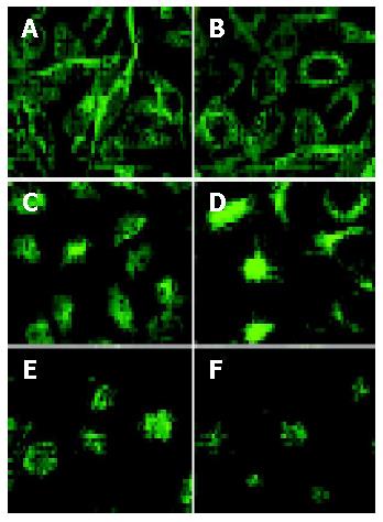
Cells co-cultured with VacA-BCS for 48 h showed the normal microtubules, which dispersed in a radial fashion throughout the cytosol, while those co-cultured with VacA+ BCS for 24 h showed collapse and disruption of the microtubule cytoarchitecture. Cells co-cultured with VacA+ BCS for 48 h had a lower density of microtubular fibers and even partially disappeared (Figure 1).

Figure 1
Figure 1 Effects of H pylori culture supernatant containing VacA on cytoskeleton of HeLa and SGC7901 cells: HeLa and SGC7901 cells co-cultured with VacA-BCS for 48 h (panels A and B) and 24 h (panels C and D), showed normal microtubules dispersed in a radial fashion throughout the cytosol, HeLa and SGC7901 cells co-cultured with VacA+BCS for 24 h (panels C and D) showed collapse and disruption of the microtubule cytoarchitecture. HeLa and SGC790 cells co-cultured with VacA+BCS for 48 h (panels E and F), showed a lower density of microtubular fibers and even partially disappeared.
DISCUSSION

The cDNA micoarray technology can screen thousands of genes responsive to some virulence determinants simultaneously and identify host gene expression patterns, during which the relationship between a virulence determinant and regulated genes of host cells may be established. It was used in the present study to examine the transcriptional response of gastric epithelial SGC7901 cells to VacA BCS in vitro.

H pylori infection could result in various diseases such as chronic gastritis, peptic ulcer and gastric carcinoma[4]. It is helpful to understand the biological process and the pathogenesis of H pylori-associated diseases and the role of VacA in the pathogenicity of H pylori by finding out gene expression alterations in cells challenged by H pylori. In this study, target cells were always incubated with H pylori or its components to observe the biological changes. After co-incubation, target cells often had growth arrest, vacuolation, apoptosis, cytokine secretion, and cytoskeleton rearrangement[5-8]. Gene chip technique was used to clarify the biological response of cells to H pylori or its toxic components and the mechanism of H pylori-related diseases. The results showed that there were 198 genes with 3 fold different expression levels and 865 genes with 2 fold different expression levels in cells challenged by VacA+ BCS. Genes with a distinct expression difference account for about 5% of total genes[9-11] and those with a definite function are involved in many important cell processes. These genes include oncogene, tumor suppressor gene, ion channel transportation associated gene, gene associated with stress response and inflammation, cell cycle-related gene, cytoskeleton-associated gene and gene related to cell apoptosis.

Expression alteration of cytoskeleton associated genes

VacA is able to delay healing of gastric ulcer in rats, inhibit reepithelialization, and worsen the quality of mucosa scar in vivo[12], inhibit in vitro gastric epithelial cell proliferation and interfere with EGF-induced signal transduction essential for healing of gastric mucosa[13]. VacA could disrupt cytoskeletal architecture essential for the maintenance of cell structure integrity and epithelial barrier function[14]. Studies by Ashorn et al[15] favored the role of VacA in cytoskeletal rearrangements, because cytotoxin-produed H pylori strains could lead to condensation of filamentous actin, while cytotoxin-negative strains did not cause marked disturbances in the intracellular structure of cells. Cell vacuolation is associated with cytoskeletal rearrangements[16,17], VacA-mediated cell vacuolation is strongly correlated to the inhibition of reepithelialization and loss of stress fibers[18].

Cytoskeleton is composed of microfilaments, microtubules, and intermediate filament cross-linked bundles, which are anchored to other cellular components, including cell membrane. Microtubules are important cytoplasmic structures involved in intracellular transport and are essential for cell division and differentiation. Intermediate filaments consist of fibrous protein, which has been shown to have mechanical functions in stiffening cells and organizing intracellular organelles for coordinated activity[14]. Our microarray analysis indicates cytoskeleton-related genes are expressed at low levels, including kinesin-like-5 protein, actin-related protein 2 (ARP2), intermediate filament protein syncoilin and dynactin 4 (Table 1). Kinesin-like-5 protein is a member of the kinesin-like protein superfamily, specifically regulates microtubule formation and consecutive movement of chromosomes during mitosis, and therefore is crucial for cell division. In recent years, actin-related protein 2/3 (Arp2/3) complex has emerged as a central effector of actin assembly that receives multiple signal inputs inducing the formation of branched filaments. The Arp2/3 complex is composed of 7 evolutionarily conserved subunits: 2 actin-related proteins (Arp2 and Arp3) and 5 other subunits, which in yeast are called Arc40, Arc35, Arc18, Arc19, and Arc15. In all organisms examined, the Arp2/3 complex is localized at the site of dynamic actin assembly. In yeast, the Arp2/3 complex is localized at the cortical actin patches, highly motile filamentous actin structures. Mutations in different subunits of the yeast Arp2/3 complex could disrupt actin organization, actin patch motility, and actin-dependent processes such as endocytosis, cell polarity development, and organelle inheritance[19]. Syncoilin is a 64-kDa protein found in skeletal and cardiac muscles and has been proposed as a member of the intermediate filament (IF) protein superfamily based on sequence analysis. Syncoilin is involved in the anchoring of the desmin intermediate filament network at the sarcolemma and neuromuscular junction that is likely to be important in maintaining muscle fiber integrity and may also link dystrophin-associated protein complex to cytoskeleton. Its dysfunction or absence might result in disruption of the intermediate filament network and muscle necrosis[20]. Dynactin is a complex with at least 10 polypeptides from 24 to 150 ku in size. The best characterized subunits of dynactin are p150Glued and p50 (dynamitin). p150Glued contains both microtubule- and dynein-binding domains. p150Glued could bind to microtubules through the NH2-terminal CAP-Gly motif, and phosphorylation near this motif has been shown to regulate microtubule binding. p150Glued could interact directly with the intermediate chain of cytoplasmic dynein (DIC) that may be essential for dynein-mediated organelle transport. Regulation of this interaction by DIC phosphorylation has suggested an important function[21]. Expression of genes encoding cytoskeleton-associated proteins essential in keeping the normal cell architecture is inhibited after cells are challenged by VacA+ BCS. This decrease results in reduction of the production of proteins encoded by these genes and collapse of cytoarchitecture. This alteration of gene expression is an early response of cells interfered with exotic factors, which precedes morphologic change of cell configuration and is the fundamental cause of cell morphologic change. But further study is still needed to clarify the mechanism of VacA+ BCS to induce cytoskeleton changes and the relationship between morphologic and cytoskeleton changes of cells. Further research on the effect of VacA+ BCS on cell morphology especially on cytoskeleton will be helpful to understand how VacA and other secreted bacterial toxic factors induce cell pathological changes.

Expression change of tumorigenesis-associated genes

H pylori is associated with gastric cancer and MALT-lymphoma. In 1994, H pylori was classified by the International Association for Research against Cancer as a type 1 carcinogen[22]. In this study, some tumor-associated genes were highly expressed, such as melanoma antigen (MAGE) gene, jun B oncogene, homologue of mouse MAT-1 oncogene, annexin A10 gene, telomeric repeat binding factor 2 gene, fibroblast growth factor receptor gene, S phase kinase associated protein (p45) gene. MAGE exists in tumor cells but not in normal cells, and is related to differentiation of tumor cells. It is a symbol of malignant cells[23,24]. Expression of tumor suppressor gene that encodes KIAA0456 protein (Dab2) decreases in 80% oophoro carcinomas. Inactivation or down-expression of Dab2 could probably induce or promote the development of oophoro carcinoma[25]. Our results show that tumor suppressor genes such as Dab2 were up-regulated, which is inconsistent with the fact that VacA+ BCS could result in increased expression of most tumor associated genes. But we found the same thing from other correlative studies, in which the expression level of tumor suppressor genes increased correspondingly with increased expression levels of oncogenes. Several ANXs are involved in tumorigenesis. ANXA1 is overexpressed in breast cancer and hepatocellular carcinoma (HCC). ANXA2 is overexpressed in brain glial tumors and pancreatic cancer and ANXA8 in acute promyelocytic leukemia. Overexpression of ANXA1 correlates positively with metastatic potential of breast cancer and increased growth of tumor cells inoculated in nude mice. In contrast, ANXA6 has tumor suppressive activities in squamous cell carcinoma, and reduction of ANXA6 and ANXA7 proteins is associated with malignant phenotype and the metastatic potential of melanoma. Moreover, ANXA4 plays a role in chemoresistance. ANXA10 may be a potential tumor suppressor gene because its down-regulation is associated with malignant phenotype of liver cells, and vascular invasion and progression of HCC[26]. Proliferation and death are two different physiological behaviors of cells. The equilibration between them is helpful to keep cell stabilization. Disruption of this equilibration would induce cell pathologic changes such as apoptosis or proliferation. VacA+ BCS could enhance these two important potentials of cells, and consequently boost the sensitivity of cells to pathogens.

Telomerase, a reverse transcriptase composed of RNA and proteins, can synthesize telomeric repeat sequence and add this sequence to the terminal of chromosome to keep the eternal life of cells. Activation of telomerase is a biological characteristic of malignant tumor[27]. In this study, increased expression of TP-1, the main component of telomerase, was observed. The expression state of tolemerase in cells challenged by VacA+ BCS still needs to be confirmed. The expression alterations of tumor-related genes in cells challenged by VacA+ BCS indicates that VacA+ BCS has the potential to promote canceration of cells.

Expression alterations of stress response genes and genes encoding inflammation-associated factors

Stress or heat shock proteins (HSPs) are ubiquitous and highly conserved proteins whose expression is induced in response to a wide variety of physiological and environmental insults. They allow cells to survive in otherwise lethal conditions. Some of the important house-keeping functions attributable to the molecular chaperones include: import of proteins into cellular compartments; folding of proteins in cytosol, endoplasmic reticulum, and mitochondria; degradation of unstable proteins; dissolution of protein complexes; prevention of protein aggregation; control of regulatory proteins and refolding of misfolded proteins[28]. Accumulation of abnormally folded proteins in nuclei or cytosol that occurs as a consequence of stress, increases temperature, free oxygen radicals, heavy metals, and even antibiotics[29]. Formation of aggregates could disturb normal cellular function and trigger cell death. Such a mechanism is involved in the pathogenesis of lesions that characterize several neurodegenerative disorders. Most HSPs are involved in the proper folding and/or elimination of misfolded proteins, thus contributing to cell survival. The relationship between HSPs and tumors is reported[30]. The yield of HSPs is increased in canceration and tumor cells[31]. In this study, expression of all HSP genes increased, indicating that abnormal states of target cells and the potential of VacA+ BCS can induce cell canceration.

IL-14 secreted by activated T and B cells can activate division of B cells and promote proliferation of some subgroups of B cells[32]. In this study, decreased expression of IL-14 gene was found to be different from other inflammatory factors. Gene expression level of other inflammation-associated factors increased, such as α-interferon, tumor necrosis factor receptor- associated proteins, interlukin-1 receptor antagonist. These results are coincident with the cell response to H pylori and its metabolic products. The interfering factor in this study was VacA+ BCS. Thus we could deduce that VacA might play an important role in this process.

The results of this study clearly indicate that H pylori VacA+ BCSs can induce expression changes at multi-gene level in infected cells, thus adjusting the expressed products according to the interference with various physiological functions and activities of target cells.

In conclusion, VacA+ BCS can cause alterations of cell gene expression profile at multi-level and multi-phase and such alterations are almost involved in all important pathologic processes of H pylori-associated diseases. It is suggested that VacA plays an important role in the pathogenic mechanism of H pylori.

Footnotes

Edited by Wang XL and Ren SY

References
1.  Ogura K, Maeda S, Nakao M, Watanabe T, Tada M, Kyutoku T, Yoshida H, Shiratori Y, Omata M. Virulence factors of Helicobacter pylori responsible for gastric diseases in Mongolian gerbil. J Exp Med. 2000;192:1601-1610.  [PubMed]  [DOI]  [Cited in This Article: ]  [Cited by in Crossref: 224]  [Cited by in F6Publishing: 224]  [Article Influence: 9.0]  [Reference Citation Analysis (0)]
2.  Leunk RD, Johnson PT, David BC, Kraft WG, Morgan DR. Cytotoxic activity in broth-culture filtrates of Campylobacter pylori. J Med Microbiol. 1988;26:93-99.  [PubMed]  [DOI]  [Cited in This Article: ]  [Cited by in Crossref: 488]  [Cited by in F6Publishing: 430]  [Article Influence: 11.6]  [Reference Citation Analysis (0)]
3.  Sepulveda AR. Molecular testing of Helicobacter pylori-associated chronic gastritis and premalignant gastric lesions: clinical implications. J Clin Gastroenterol. 2001;32:377-382.  [PubMed]  [DOI]  [Cited in This Article: ]  [Cited by in Crossref: 12]  [Cited by in F6Publishing: 12]  [Article Influence: 0.5]  [Reference Citation Analysis (0)]
4.  Mann NS, Westblom TU. Helicobacter pylori and the future: an afterword. Curr Top Microbiol Immunol. 1999;241:301-308.  [PubMed]  [DOI]  [Cited in This Article: ]  [Cited by in Crossref: 3]  [Cited by in F6Publishing: 5]  [Article Influence: 0.2]  [Reference Citation Analysis (0)]
5.  Ogura K, Takahashi M, Maeda S, Ikenoue T, Kanai F, Yoshida H, Shiratori Y, Mori K, Mafune KI, Omata M. Interleukin-8 production in primary cultures of human gastric epithelial cells induced by Helicobacter pylori. Dig Dis Sci. 1998;43:2738-2743.  [PubMed]  [DOI]  [Cited in This Article: ]  [Cited by in Crossref: 25]  [Cited by in F6Publishing: 28]  [Article Influence: 1.0]  [Reference Citation Analysis (0)]
6.  Wagner S, Beil W, Westermann J, Logan RP, Bock CT, Trautwein C, Bleck JS, Manns MP. Regulation of gastric epithelial cell growth by Helicobacter pylori: offdence for a major role of apoptosis. Gastroenterology. 1997;113:1836-1847.  [PubMed]  [DOI]  [Cited in This Article: ]  [Cited by in Crossref: 222]  [Cited by in F6Publishing: 236]  [Article Influence: 8.4]  [Reference Citation Analysis (0)]
7.  Cover TL, Halter SA, Blaser MJ. Characterization of HeLa cell vacuoles induced by Helicobacter pylori broth culture supernatant. Hum Pathol. 1992;23:1004-1010.  [PubMed]  [DOI]  [Cited in This Article: ]  [Cited by in Crossref: 54]  [Cited by in F6Publishing: 54]  [Article Influence: 1.6]  [Reference Citation Analysis (0)]
8.  Segal ED, Falkow S, Tompkins LS. Helicobacter pylori attachment to gastric cells induces cytoskeletal rearrangements and tyrosine phosphorylation of host cell proteins. Proc Natl Acad Sci USA. 1996;93:1259-1264.  [PubMed]  [DOI]  [Cited in This Article: ]  [Cited by in Crossref: 204]  [Cited by in F6Publishing: 215]  [Article Influence: 7.4]  [Reference Citation Analysis (0)]
9.  Cox JM, Clayton CL, Tomita T, Wallace DM, Robinson PA, Crabtree JE. cDNA array analysis of cag pathogenicity island-associated Helicobacter pylori epithelial cell response genes. Infect Immun. 2001;69:6970-6980.  [PubMed]  [DOI]  [Cited in This Article: ]  [Cited by in Crossref: 74]  [Cited by in F6Publishing: 78]  [Article Influence: 3.3]  [Reference Citation Analysis (0)]
10.  Chiou CC, Chan CC, Sheu DL, Chen KT, Li YS, Chan EC. Helicobacter pylori infection induced alteration of gene expression in human gastric cells. Gut. 2001;48:598-604.  [PubMed]  [DOI]  [Cited in This Article: ]  [Cited by in Crossref: 38]  [Cited by in F6Publishing: 41]  [Article Influence: 1.7]  [Reference Citation Analysis (0)]
11.  Maeda S, Otsuka M, Hirata Y, Mitsuno Y, Yoshida H, Shiratori Y, Masuho Y, Muramatsu M, Seki N, Omata M. cDNA microarray analysis of Helicobacter pylori-mediated alteration of gene expression in gastric cancer cells. Biochem Biophys Res Commun. 2001;284:443-449.  [PubMed]  [DOI]  [Cited in This Article: ]  [Cited by in Crossref: 65]  [Cited by in F6Publishing: 70]  [Article Influence: 2.9]  [Reference Citation Analysis (0)]
12.  Chang K, Fujiwara Y, Wyle F, Tarnawski A. Helicobacter pylori toxin inhibits growth and proliferation of cultured gastric cells-Kato III. J Physiol Pharmacol. 1993;44:17-22.  [PubMed]  [DOI]  [Cited in This Article: ]
13.  Fujiwara Y, Wyle F, Arakawa T, Domek MJ, Fukuda T, Kobayashi K, Tarnawski A. Helicobacter pylori culture supernatant inhibits binding and proliferative response of human gastric cells to epidermal growth factor: implications for H.pylori interference with ulcer healing? Digestion. 1997;58:299-303.  [PubMed]  [DOI]  [Cited in This Article: ]  [Cited by in Crossref: 15]  [Cited by in F6Publishing: 17]  [Article Influence: 0.6]  [Reference Citation Analysis (0)]
14.  Pai R, Cover TL, Tarnawski AS. Helicobacter pylori vacuolating cytotoxin (VacA) disorganizes the cytoskeletal architecture of gastric epithelial cells. Biochem Biophys Res Commun. 1999;262:245-250.  [PubMed]  [DOI]  [Cited in This Article: ]  [Cited by in Crossref: 41]  [Cited by in F6Publishing: 37]  [Article Influence: 1.4]  [Reference Citation Analysis (0)]
15.  Ashorn M, Cantet F, Mayo K, Megraud F. Cytoskeletal rearrangements induced by Helicobacter pylori strains in epithelial cell culture: possible role of the cytotoxin. Dig Dis Sci. 2000;45:1774-1780.  [PubMed]  [DOI]  [Cited in This Article: ]  [Cited by in Crossref: 9]  [Cited by in F6Publishing: 10]  [Article Influence: 0.4]  [Reference Citation Analysis (0)]
16.  Henics T, Wheatley DN. Vacuolar cytoplasmic phase separation in cultured mammalian cells involves the microfilament network and reduces motional properties of intracellular water. Int J Exp Pathol. 1997;78:343-354.  [PubMed]  [DOI]  [Cited in This Article: ]  [Cited by in Crossref: 9]  [Cited by in F6Publishing: 9]  [Article Influence: 0.3]  [Reference Citation Analysis (0)]
17.  Henics T, Wheatley DN. Cytoplasmic vacuolation, adaptation and cell death: a view on new perspectives and features. Biol Cell. 1999;91:485-498.  [PubMed]  [DOI]  [Cited in This Article: ]  [Cited by in Crossref: 138]  [Cited by in F6Publishing: 141]  [Article Influence: 5.4]  [Reference Citation Analysis (0)]
18.  Pai R, Sasaki E, Tarnawski AS. Helicobacter pylori vacuolating cytotoxin (VacA) alters cytoskeleton-associated proteins and interferes with re-epithelialization of wounded gastric epithelial monolayers. Cell Biol Int. 2000;24:291-301.  [PubMed]  [DOI]  [Cited in This Article: ]  [Cited by in Crossref: 19]  [Cited by in F6Publishing: 17]  [Article Influence: 0.7]  [Reference Citation Analysis (0)]
19.  Humphries CL, Balcer HI, D'Agostino JL, Winsor B, Drubin DG, Barnes G, Andrews BJ, Goode BL. Direct regulation of Arp2/3 complex activity and function by the actin binding protein coronin. J Cell Biol. 2002;159:993-1004.  [PubMed]  [DOI]  [Cited in This Article: ]  [Cited by in Crossref: 163]  [Cited by in F6Publishing: 165]  [Article Influence: 7.2]  [Reference Citation Analysis (0)]
20.  Poon E, Howman EV, Newey SE, Davies KE. Association of syncoilin and desmin: linking intermediate filament proteins to the dystrophin-associated protein complex. J Biol Chem. 2002;277:3433-3439.  [PubMed]  [DOI]  [Cited in This Article: ]  [Cited by in Crossref: 64]  [Cited by in F6Publishing: 62]  [Article Influence: 2.7]  [Reference Citation Analysis (0)]
21.  Deacon SW, Serpinskaya AS, Vaughan PS, Lopez Fanarraga M, Vernos I, Vaughan KT, Gelfand VI. Dynactin is required for bidirectional organelle transport. J Cell Biol. 2003;160:297-301.  [PubMed]  [DOI]  [Cited in This Article: ]  [Cited by in Crossref: 255]  [Cited by in F6Publishing: 265]  [Article Influence: 12.0]  [Reference Citation Analysis (0)]
22.  IARC working group on the evaluation of carcinogenic risks to humans: some industrial chemicals. Lyon, 15-22 February 1994. IARC Monogr Eval Carcinog Risks Hum. 1994;60:1-560.  [PubMed]  [DOI]  [Cited in This Article: ]
23.  Park JH, Lee SW. Hypertonicity induction of melanoma antigen, a tumor-associated antigen. Mol Cells. 2002;13:288-295.  [PubMed]  [DOI]  [Cited in This Article: ]
24.  Kavalar R, Sarcevic B, Spagnoli GC, Separovic V, Samija M, Terracciano L, Heberer M, Juretic A. Expression of MAGE tumour-associated antigens is inversely correlated with tumour differentiation in invasive ductal breast cancers: an immunohistochemical study. Virchows Arch. 2001;439:127-131.  [PubMed]  [DOI]  [Cited in This Article: ]  [Cited by in Crossref: 28]  [Cited by in F6Publishing: 31]  [Article Influence: 1.3]  [Reference Citation Analysis (0)]
25.  Yang DH, Smith ER, Cohen C, Wu H, Patriotis C, Godwin AK, Hamilton TC, Xu XX. Molecular events associated with dysplastic morphologic transformation and initiation of ovarian tumorigenicity. Cancer. 2002;94:2380-2392.  [PubMed]  [DOI]  [Cited in This Article: ]  [Cited by in Crossref: 59]  [Cited by in F6Publishing: 63]  [Article Influence: 2.7]  [Reference Citation Analysis (0)]
26.  Liu SH, Lin CY, Peng SY, Jeng YM, Pan HW, Lai PL, Liu CL, Hsu HC. Down-regulation of annexin A10 in hepatocellular carcinoma is associated with vascular invasion, early recurrence, and poor prognosis in synergy with p53 mutation. Am J Pathol. 2002;160:1831-1837.  [PubMed]  [DOI]  [Cited in This Article: ]  [Cited by in Crossref: 61]  [Cited by in F6Publishing: 71]  [Article Influence: 3.1]  [Reference Citation Analysis (0)]
27.  Hiyama E, Hiyama K. Telomerase as tumor marker. Cancer Lett. 2003;194:221-233.  [PubMed]  [DOI]  [Cited in This Article: ]  [Cited by in Crossref: 144]  [Cited by in F6Publishing: 125]  [Article Influence: 5.7]  [Reference Citation Analysis (0)]
28.  Bukau B, Horwich AL. The Hsp70 and Hsp60 chaperone machines. Cell. 1998;92:351-366.  [PubMed]  [DOI]  [Cited in This Article: ]  [Cited by in Crossref: 2149]  [Cited by in F6Publishing: 2049]  [Article Influence: 75.9]  [Reference Citation Analysis (0)]
29.  Sherman MY, Goldberg AL. Cellular defenses against unfolded proteins: a cell biologist thinks about neurodegenerative diseases. Neuron. 2001;29:15-32.  [PubMed]  [DOI]  [Cited in This Article: ]  [Cited by in Crossref: 758]  [Cited by in F6Publishing: 729]  [Article Influence: 30.4]  [Reference Citation Analysis (0)]
30.  Fałkowska-Podstawka M, Wernicki A. Heat shock proteins in health and disease. Pol J Vet Sci. 2003;6:61-70.  [PubMed]  [DOI]  [Cited in This Article: ]
31.  Hickey E, Brandon SE, Smale G, Lloyd D, Weber LA. Sequence and regulation of a gene encoding a human 89-kilodalton heat shock protein. Mol Cell Biol. 1989;9:2615-2626.  [PubMed]  [DOI]  [Cited in This Article: ]
32.  Ambrus JL, Fauci AS. Human B lymphoma cell line producing B cell growth factor. J Clin Invest. 1985;75:732-739.  [PubMed]  [DOI]  [Cited in This Article: ]  [Cited by in Crossref: 77]  [Cited by in F6Publishing: 90]  [Article Influence: 2.3]  [Reference Citation Analysis (0)]