©The Author(s) 2003.
World J Gastroenterol. Jul 15, 2003; 9(7): 1404-1408
Published online Jul 15, 2003. doi: 10.3748/wjg.v9.i7.1404
Published online Jul 15, 2003. doi: 10.3748/wjg.v9.i7.1404
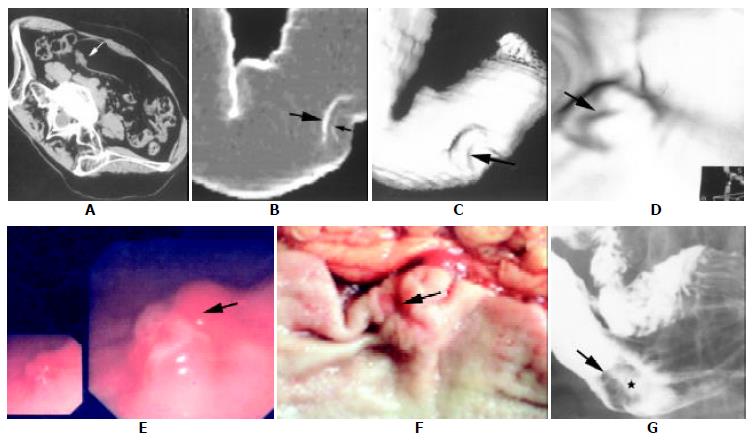
Figure 1 A complete set of image data of a patient.
Spiral CT images (A-D), fiberoptic gastroscopy (E), surgical specimen (F) and corresponding barium study (G) from a patient (female, 48 a.) of Borrmann's type 2 and TNM stage 1 of advanced gastric carcinoma. (A), axial image (supine position with right side elevated) showed a focal irregular protruding (arrow) from the posterior wall of antrum. (B), "Raysum"display (virtual double contrast barium study) stressed double-margin changes at the greater curvature of antrum: tumor (large arrow) and ulcer (small arrow) margins. (C), SSD image showed depression at the antrum with a central ulcer (arrow). (D), CTVG image depicted an intraluminal irregular mass with a flat ulcer (arrow). The view angle was illustrated by the 2D image at lower right corner. (E), fiberoptic gastroscopic view showed a lobulated mass.(F), surgical specimen demon stratad a mass with a small central ulcer (arrow). (G), barium study revealed an intraluminal filling defect (arrows) with a flat ulcer (asterisk) at the greater curvature of antrum.
- Citation: Chen F, Ni YC, Zheng KE, Ju SH, Sun J, Ou XL, Xu MH, Zhang H, Marchal G. Spiral CT in gastric carcinoma: Comparison with barium study, fiberoptic gastroscopy and histopathology. World J Gastroenterol 2003; 9(7): 1404-1408
- URL: https://www.wjgnet.com/1007-9327/full/v9/i7/1404.htm
- DOI: https://dx.doi.org/10.3748/wjg.v9.i7.1404


