Copyright
©The Author(s) 2003.
World J Gastroenterol. Apr 15, 2003; 9(4): 851-857
Published online Apr 15, 2003. doi: 10.3748/wjg.v9.i4.851
Published online Apr 15, 2003. doi: 10.3748/wjg.v9.i4.851
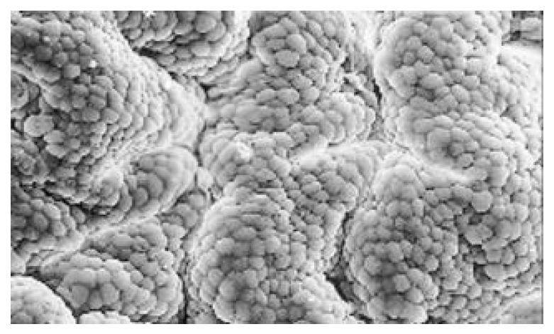
Figure 1 Convolution shape of mucosa surface.
× 320.
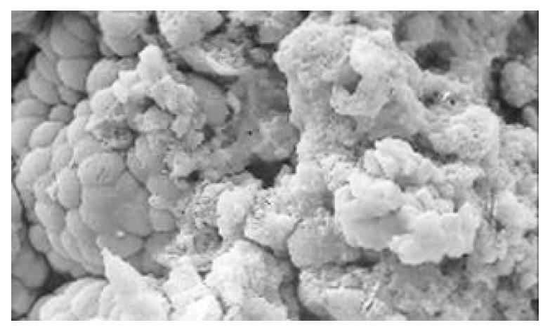
Figure 2 Degeneration, diabrosis, necrosis and exfoliation of gastric mucosa epithelial cells.
× 640.
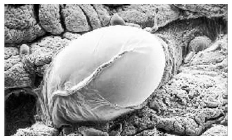
Figure 3 Cysts of gastric mucosa.
× 64.
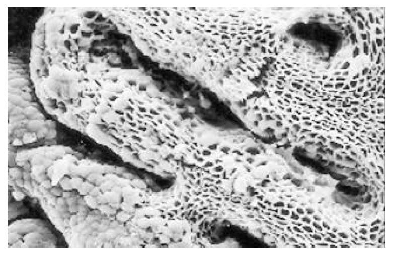
Figure 4 Atrophic and denatured glands propria in grid frame-work structure.
× 320.
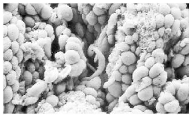
Figure 5 The surface of IM cells is thickly coated, intercellular boundaries are not clear.
× 1250.
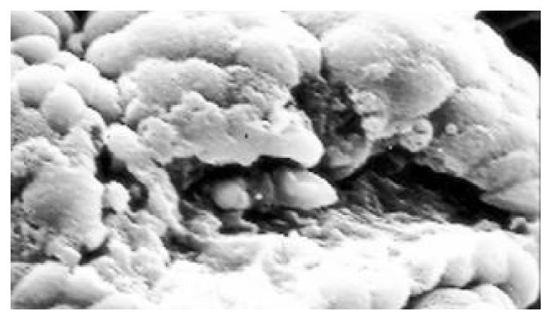
Figure 6 The hyperplastic cells are of different sizes and states, the active membranes are shaped like lesser tubercles.
× 320.
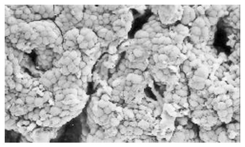
Figure 7 The hyperplastic cells are of different sizes and states, the active membranes are shaped like lesser tubercles.
× 640.
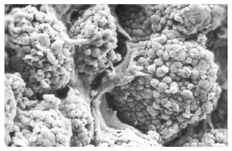
Figure 8 The cells of gastric cancer are conglobated and shaped like grapes.
× 320.
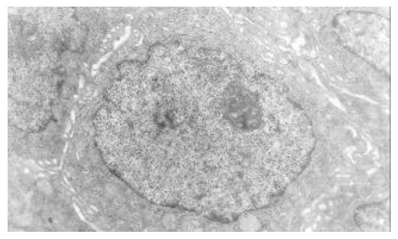
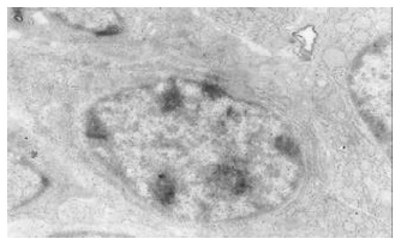
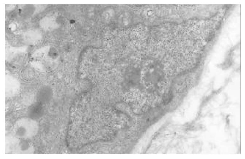
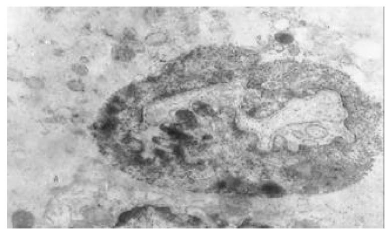
Figure 9 There is an increase in inter chromatinic grandules densification, nucleolar grandules become thick nucleoli ex-pand with irregular margins.
× 14000.
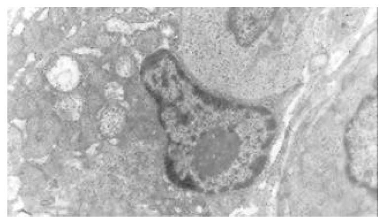
Figure 10 Nucleolar margination.
× 7200.
Figure 11 Multi nucleoli.
× 14000
Figure 12 Nucleolar division.
× 14000.
Figure 13 Intranuclear inclusion.
× 14000.
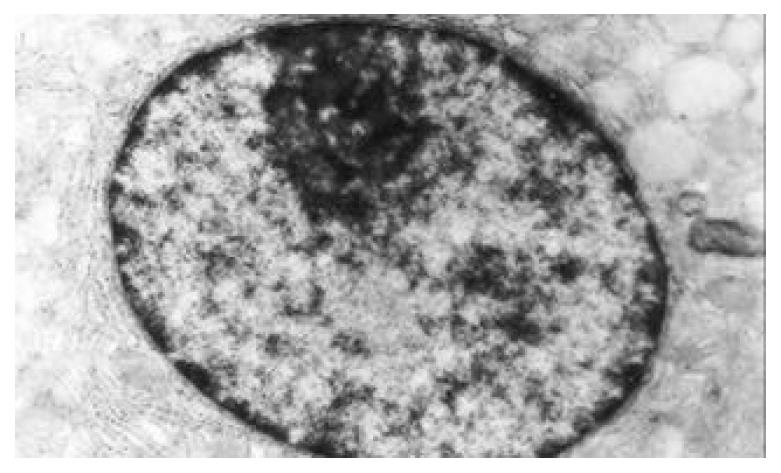
Figure 14 The densification of inter chromatinic grandules.
× 14000.
- Citation: Yin GY, Zhang WN, Shen XJ, Chen Y, He XF. Ultrastructure and molecular biological changes of chronic gastritis, gastric cancer and gastric precancerous lesions: a comparative study. World J Gastroenterol 2003; 9(4): 851-857
- URL: https://www.wjgnet.com/1007-9327/full/v9/i4/851.htm
- DOI: https://dx.doi.org/10.3748/wjg.v9.i4.851


