©The Author(s) 2003.
World J Gastroenterol. Apr 15, 2003; 9(4): 696-700
Published online Apr 15, 2003. doi: 10.3748/wjg.v9.i4.696
Published online Apr 15, 2003. doi: 10.3748/wjg.v9.i4.696
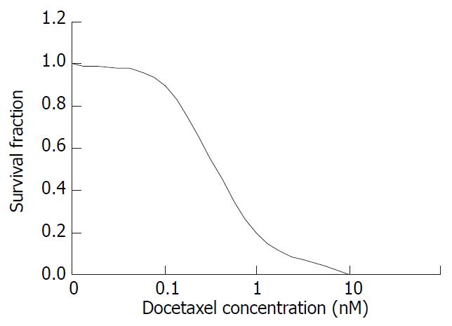
Figure 1 The survival fraction of SMMC-7721 cells treated with docetaxel for 24 hr.
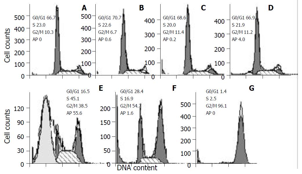
Figure 2 Cell cycle changes and apoptosis induction in SMMC-7721 cells treated with docetaxel for 24hr.
Cells were treated with docetaxel at 0 M (A), 10-11 M (B), 10-10 M (C), 10-9 M (D), 10-8 M (E), 10-7 M (F), 10-6 M (G). A marked apoptosis was induced at 10-8 M, and a peaked G2/M phase accumulation was caused at 10-6 M. Ap: apoptosis.
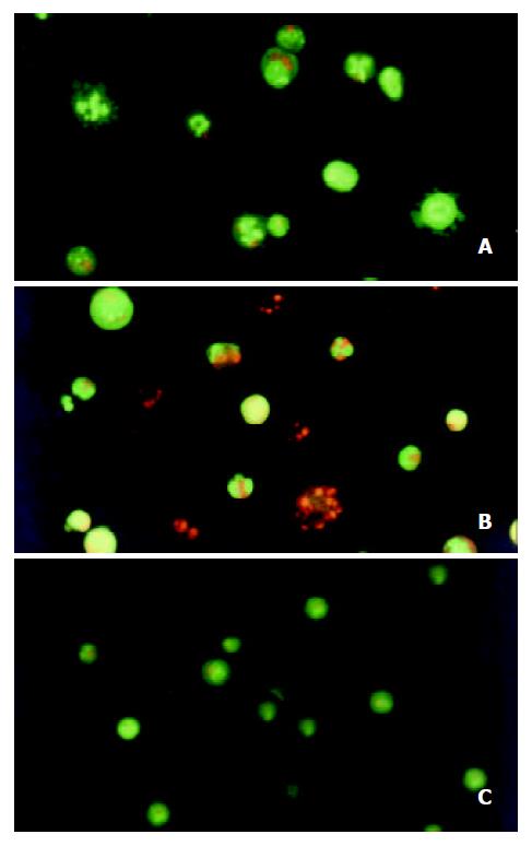
Figure 3 Morphological study with fluorescence microscope.
After the SMMC-7721 cells were given 10-8 M docetaxel for 24 hr (A), the cells were stained green with AO, the nuclei exhibited bright condensed chromatin or fragmented, some cells blebbed. After being treated for 48 hr (B), cells were stained red with EB, the apoptotic bodies could be seen clearly. On the contrary, the untreated cells (C) did not show these apoptotic characteristics.
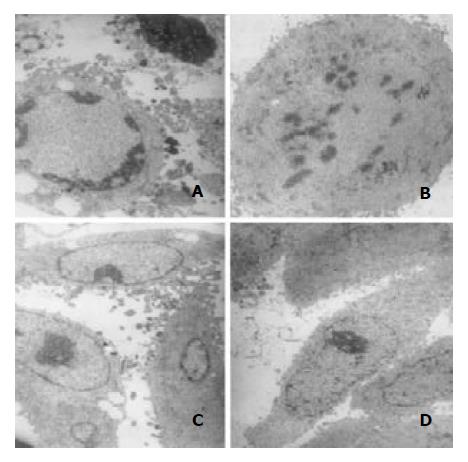
Figure 4 Transmission electron microscopic observation.
Af-ter treatment with 10-8 M docetaxel for 24 hr, the chromatin of some SMMC-7721 cells was located along the nuclear edges or formed irregularly shaped crescents at the nuclear edges (A, C),or became condensed or fragmented (A, B); The vacuole could be observed in some cytoplasm (A, C).
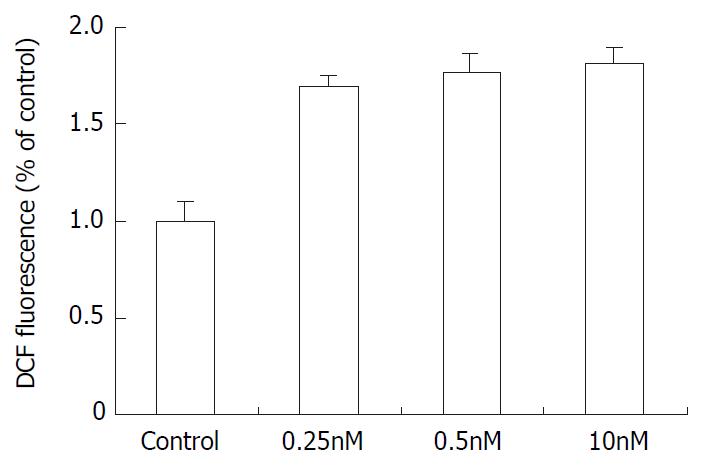
Figure 5 The effect of docetaxel on the formation of ROS in SMMC-7721 cells.
Data are from three independent experiments.
- Citation: Geng CX, Zeng ZC, Wang JY. Docetaxel inhibits SMMC-7721 human hepatocellular carcinoma cells growth and induces apoptosis. World J Gastroenterol 2003; 9(4): 696-700
- URL: https://www.wjgnet.com/1007-9327/full/v9/i4/696.htm
- DOI: https://dx.doi.org/10.3748/wjg.v9.i4.696


