©The Author(s) 2002.
World J Gastroenterol. Apr 15, 2002; 8(2): 276-281
Published online Apr 15, 2002. doi: 10.3748/wjg.v8.i2.276
Published online Apr 15, 2002. doi: 10.3748/wjg.v8.i2.276
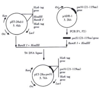
Figure 1 Construction of expression plasmid pET-28a-preS1
Math 1 Math(A1).
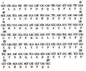
Figure 2 Nucleotide and derived amino acid sequence of PreS1 (21-119 aa) region gene fragment inserted into the expression vector pET-28a-preS1.
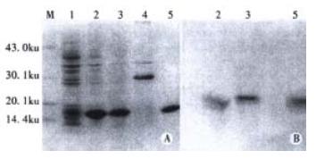
Figure 3 Expression and intracellular location of preS1(21-119 aa) in E.
coli. BL21(DE3)plysS cells. Protein samples were subjected to 15% SDS-PAGE and stained with Commassia brilliant blue (A), and the duplicate gel was electroblotted onto a nitrocellulose membrane for western analysis (B). Lane M: molecular mass standards. Lane 1-2 are culture lysates of uninduced BL21 cells (lane 1) and induced BL21 cells (lane 2). Lane 3, 4: Supernatant (soluble) and precipitate (insoluble) fractions, respectively; Lane5: purified fusion protein by Ni2+-IDA-sepharose 6B.
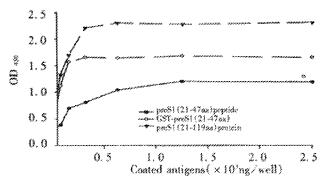
Figure 4 immunoreactivity of preS1(21-119 aa), GST-preS1 (21-47aa) peptide.
Microtiter plate was coated with different amount (10-2500 ng) of the purified preS1 proteins or peptide. Then direct ELISA was performed.
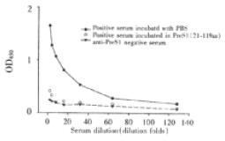
Figure 5 Specificity test of indirect ELISA for detection of anti-preS1 antibodies.
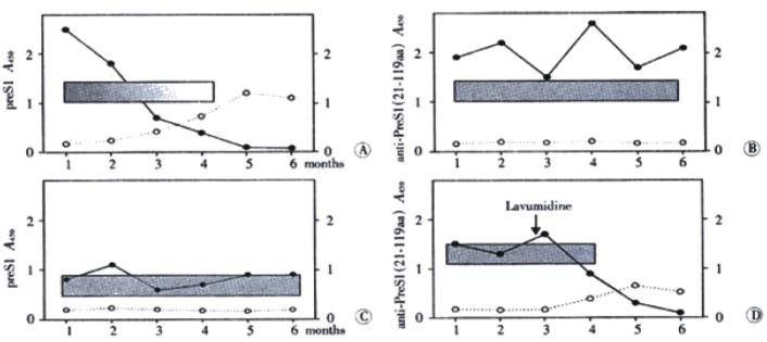
Figure 6 The profiles of serologic markers during acute chronic HBV infection with immuno-diagnosis for preS1 antigen and anti-preS1(21-119 aa) antibodies.
A: Typical serologic profile of HBV markers during acute infection, with disappearance of preS1 antigen and seroconversion to anti-preS1(21-119 aa) antibodies; elimination of HBV-DNA in 2-4 mo from the appearance of antibodies; B: Chronic patients with HBeAg+ serologic profile; high level of preS1 antigen and HBV-DNA but absence of anti-preS1(21-119 aa) antibodies; C: Chronic patients with anti-HBe+ serologic profile; low level of preS1 domain and HBV-DNA and absence of anti-preS1(21-119 aa) antibodies. D: One patient persisting of HBsAg, HBeAg and preS1 for almost 3 years until treated with Lamivudine. Appearance of anti-preS1(21-119 aa) antibodies with simultaneous health improvement; disappearance of HBV-DNA and declining level of preS1 antigen in serum. OD450 value of Y axis (‘●’ preS1 antigen and ‘○’ anti-preS1 antibody was mean value of inpatients in every group; different shade (■) in the rectangle (A) represented different level of HBV-DNA in patients.
- Citation: Wei J, Wang YQ, Lu ZM, Li GD, Wang Y, Zhang ZC. Detection of anti-preS1 antibodies for recovery of hepatitis B patients by immunoassay. World J Gastroenterol 2002; 8(2): 276-281
- URL: https://www.wjgnet.com/1007-9327/full/v8/i2/276.htm
- DOI: https://dx.doi.org/10.3748/wjg.v8.i2.276


