©The Author(s) 2001.
World J Gastroenterol. Aug 15, 2001; 7(4): 490-495
Published online Aug 15, 2001. doi: 10.3748/wjg.v7.i4.490
Published online Aug 15, 2001. doi: 10.3748/wjg.v7.i4.490
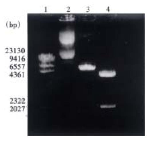
Figure 1 Identification of PCMV-Egr-1 plasmid.
1. Marker; 2. Uncut plasmid; 3. Cut with Sac II, showing 7.6 kp fragment of whole plasmid; 4. Cut with Sac II and Sma I, showing 5.5 kp fragment of vector and 2.1 kp fragment of Egr-1 gene.
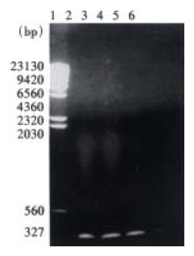
Figure 2 PCR amplification of neogene.
1. Marker; 2-5. For transfected Eca109, showing 327 bp of neo-gene; 6. Negative control Eca109, no band was shown.
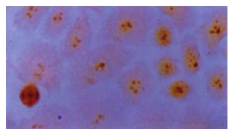
Figure 3 Positive Egr-1 protein in nuclei of transfected Eca109 cells.
ICC × 400
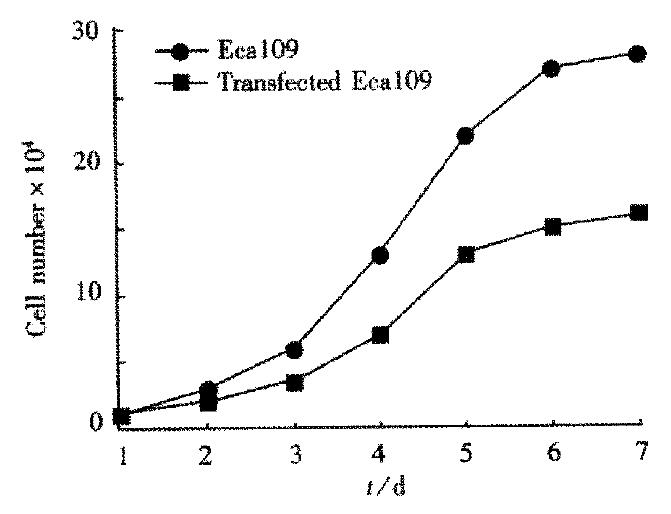
Figure 4 Cell growth curves showing lower growth rate in transfected Eca109 cell than in the control Eca109 cell.
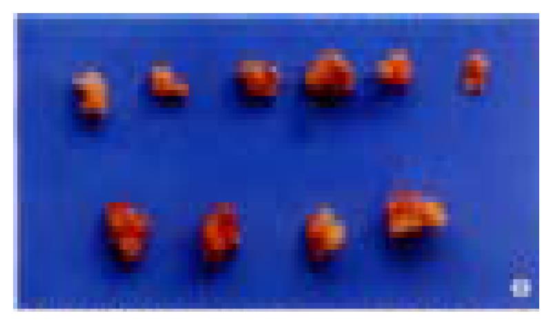
Figure 5 The tumors of transfected Eca109 are smaller than that of control Eca109, in vitro, 30 d after injection in tumorigenicity test in SCID mice.
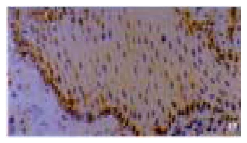
Figure 6 Egr-1 protein expression in basal mucosal layer in simple hyperplastic epithelia of esophagus.
IHC × 200
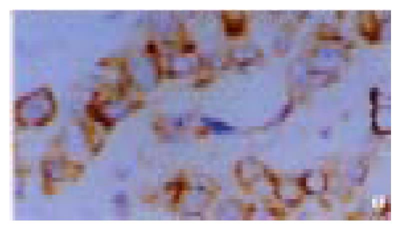
Figure 7 Positive Egr-1 mRNA in cytoplasm of esophageal squamous cell carcinoma.
ISH × 400
- Citation: Wu MY, Chen MH, Liang YR, Meng GZ, Yang HX, Zhuang CX. Experimental and clinic-opathologic study on the relationship between transcription factor Egr-1 and esophageal carcinoma. World J Gastroenterol 2001; 7(4): 490-495
- URL: https://www.wjgnet.com/1007-9327/full/v7/i4/490.htm
- DOI: https://dx.doi.org/10.3748/wjg.v7.i4.490


