Copyright
©The Author(s) 1999.
World J Gastroenterol. Jun 15, 1999; 5(3): 231-234
Published online Jun 15, 1999. doi: 10.3748/wjg.v5.i3.231
Published online Jun 15, 1999. doi: 10.3748/wjg.v5.i3.231
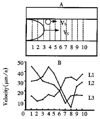
Figure 1 Leukocytes rolling along the blood vessel wall showed a “jerky” movement.
(A. Multiple sampling scheme for the velocity determination used by MIMPCAS; B. Three leukocytes passed through 10 sampling li nes with a large variation of velocity.)
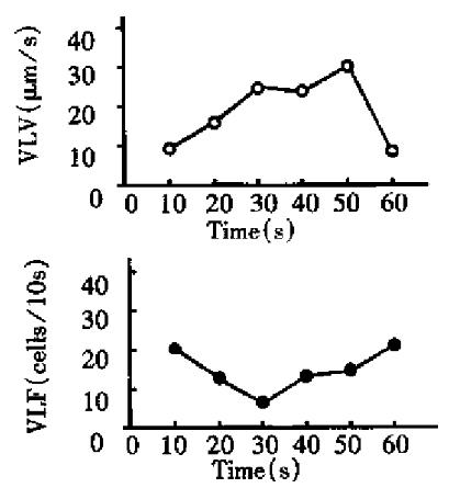
Figure 2 The time-dependent changes of VLV and VLF in the third order venules of rat mesentery.
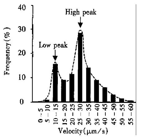
Figure 3 The frequency histogram of VLV.
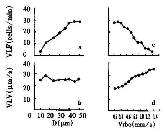
Figure 4 Effect of vessel diameter and red blood cell velocity on the flow and distribution of visible leukocytes in the venules of rats.
- Citation: Jiang Y, Liu AH, Zhao KS. Studies on the flow and distribution of leukocytes in mesentery microcirculation of rats. World J Gastroenterol 1999; 5(3): 231-234
- URL: https://www.wjgnet.com/1007-9327/full/v5/i3/231.htm
- DOI: https://dx.doi.org/10.3748/wjg.v5.i3.231


