©The Author(s) 1998.
World J Gastroenterol. Apr 15, 1998; 4(2): 128-132
Published online Apr 15, 1998. doi: 10.3748/wjg.v4.i2.128
Published online Apr 15, 1998. doi: 10.3748/wjg.v4.i2.128
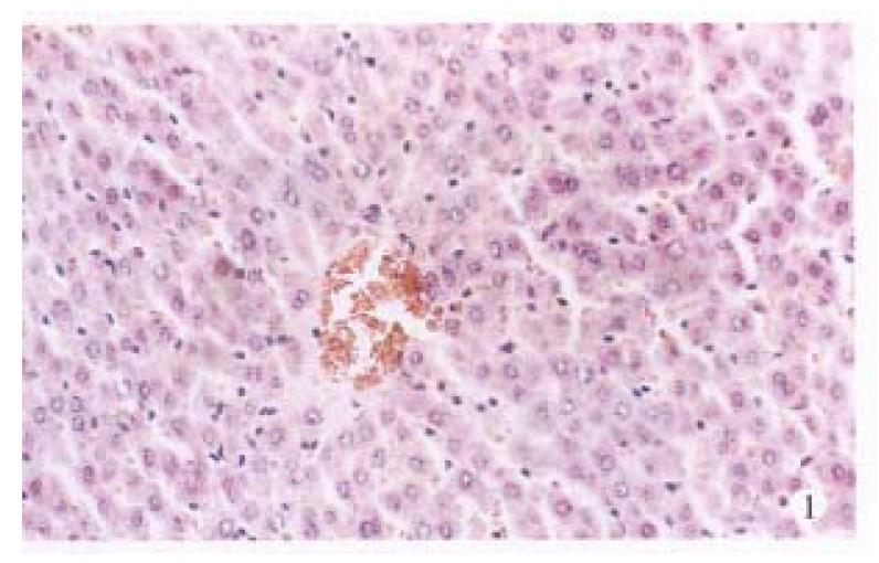
Figure 1 Section of liver from an untreated control (sham) rat.
HE × 20.
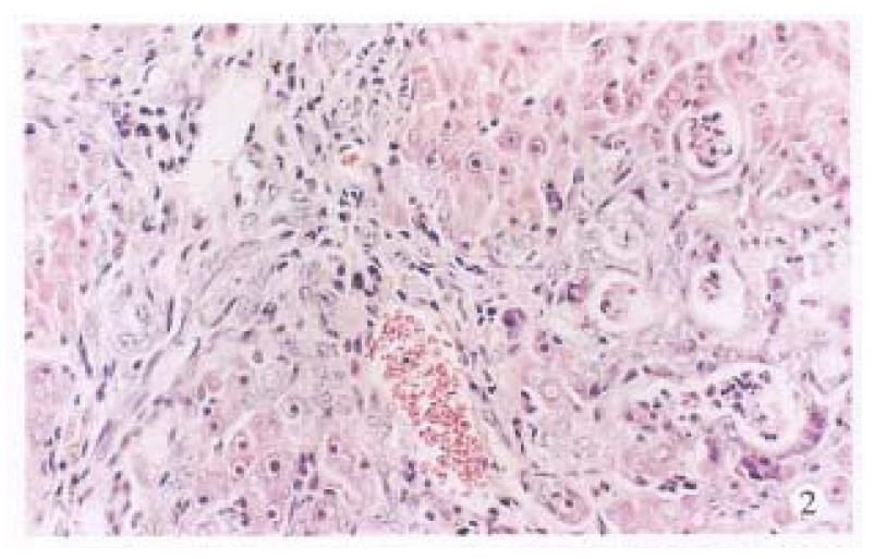
Figure 2 Section of liver from a TAA control rat treated as described in Materials and Methods.
The nodules show the transforming hepatocytes and the hepatocarcinoma. The spindle fibroblasts are recognized around such hyperplastic nodules. HE × 20.
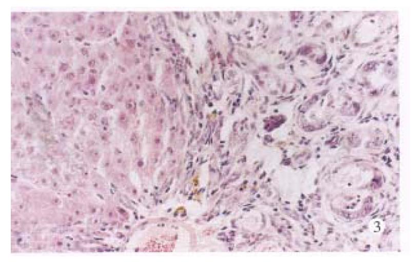
Figure 3 Section of liver from a rat in TAA + ST group.
Pseudoglandular structures and spindle cells are observed in perivenular areas. HE × 20.
Figure 4 Section of liver from a rat in TAA + ST + LPS group.
The disorganized and atypical hepatocytes formed a “mucoid lake” with floating of small cells. HE × 20.
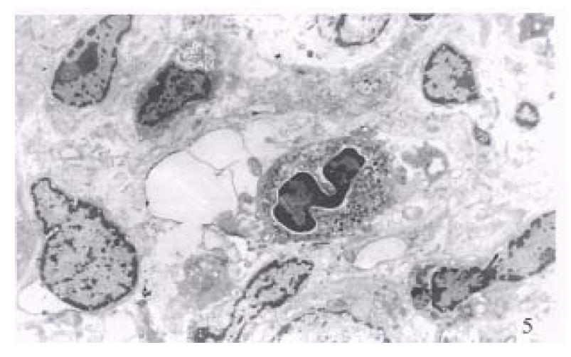
Figure 5 Ultrastructure of hepatocellular carcinoma from a rat in TAA + ST + LPS group.
Uranyl acetate and lead citrate staining × 400.
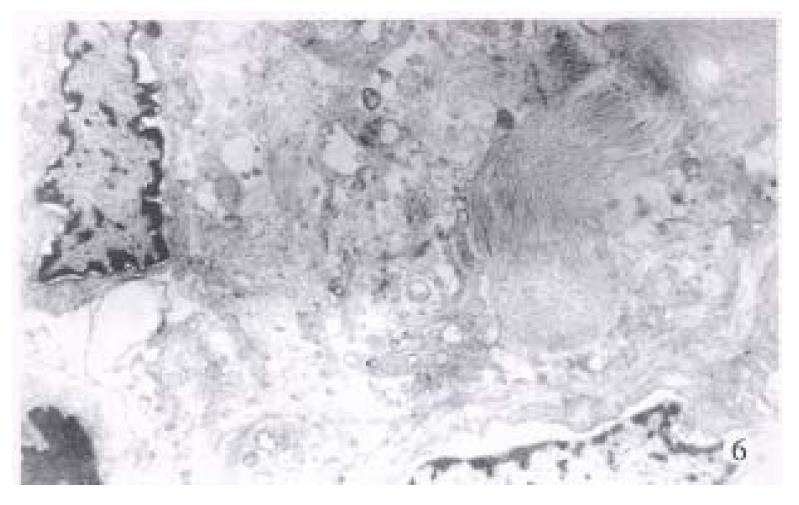
Figure 6 Ultrastructure of cirrhosis from a rat in TAA + ST + LPS group.
The bundles of collagen fibrils were present with atypical hepatocytes. Uranyl acetate and lead citrate staining × 800.
- Citation: Yang JM, Han DW, Xie CM, Liang QC, Zhao YC, Ma XH. Endotoxins enhance hepatocarcinogenesis induced by oral intake of thioacetamide in rats. World J Gastroenterol 1998; 4(2): 128-132
- URL: https://www.wjgnet.com/1007-9327/full/v4/i2/128.htm
- DOI: https://dx.doi.org/10.3748/wjg.v4.i2.128


