©The Author(s) 1998.
World J Gastroenterol. Feb 15, 1998; 4(1): 74-76
Published online Feb 15, 1998. doi: 10.3748/wjg.v4.i1.74
Published online Feb 15, 1998. doi: 10.3748/wjg.v4.i1.74
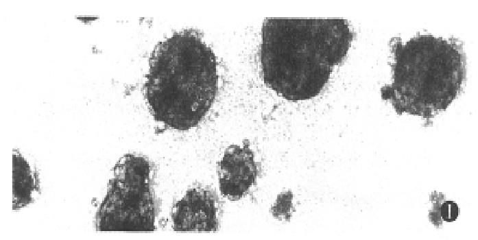
Figure 1 Phase-contrast microscopic feature of multicellular spheroidal aggregates of hepatocytes at day 4.
( × 200)
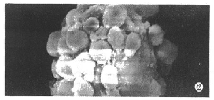
Figure 2 Scanning electron microscopy of spheroidal aggregates at day 14.
( × 1000)
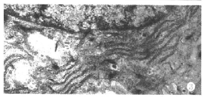
Figure 3 Transmisson electron microscopy of hepatocyte spheroids cultured for 25 days, showing abundant organelles in hepatocytes.
( × 10000)
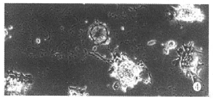
Figure 4 Phase-contrast photomicrograph of hepatocyte spheroids attached to collagen-coated wells.
( × 100)
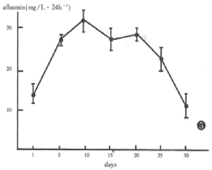
Figure 5 Albumin synthesis and secretion by aggregate hepatocytes.
- Citation: Wang YJ, Li MD, Wang YM, Ding J, Nie QH. Simplified isolation and spheroidal aggregate culture of rat hepatocytes. World J Gastroenterol 1998; 4(1): 74-76
- URL: https://www.wjgnet.com/1007-9327/full/v4/i1/74.htm
- DOI: https://dx.doi.org/10.3748/wjg.v4.i1.74


