
















Copyright: ©Author(s) 2027.
World J Gastroenterol. Jan 14, 2027; 33(2): 125661
Published online Jan 14, 2027. doi: 10.3748/wjg.125661
Published online Jan 14, 2027. doi: 10.3748/wjg.125661
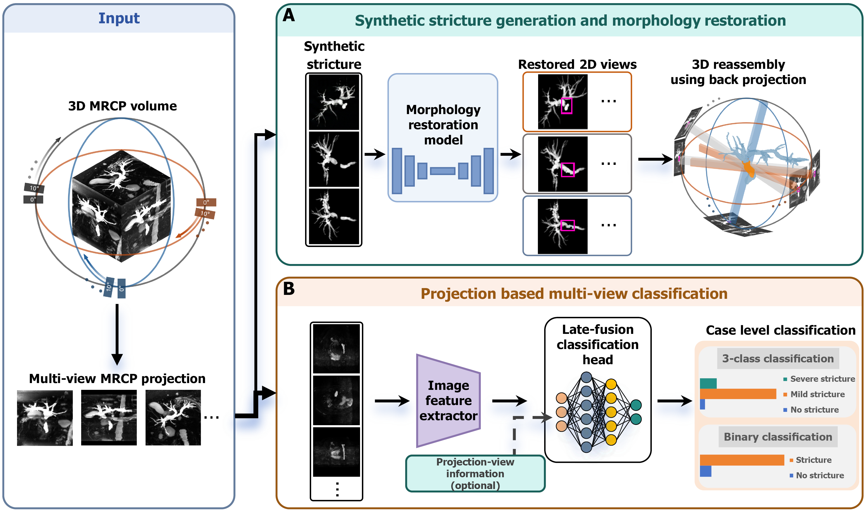
Figure 1 Magnetic resonance cholangiopancreatography-based workflow for biliary stricture reconstruction and classification.
A: Synthetic stricture generation and morphology-restoration workflow; B: Projection-based multi-view classification workflow. A shared three-dimensional magnetic resonance cholangiopancreatography-derived biliary volume was projected into multiple two-dimensional views. MRCP: Magnetic resonance cholangiopancreatography; 3D: Three-dimensional; 2D: Two-dimensional.
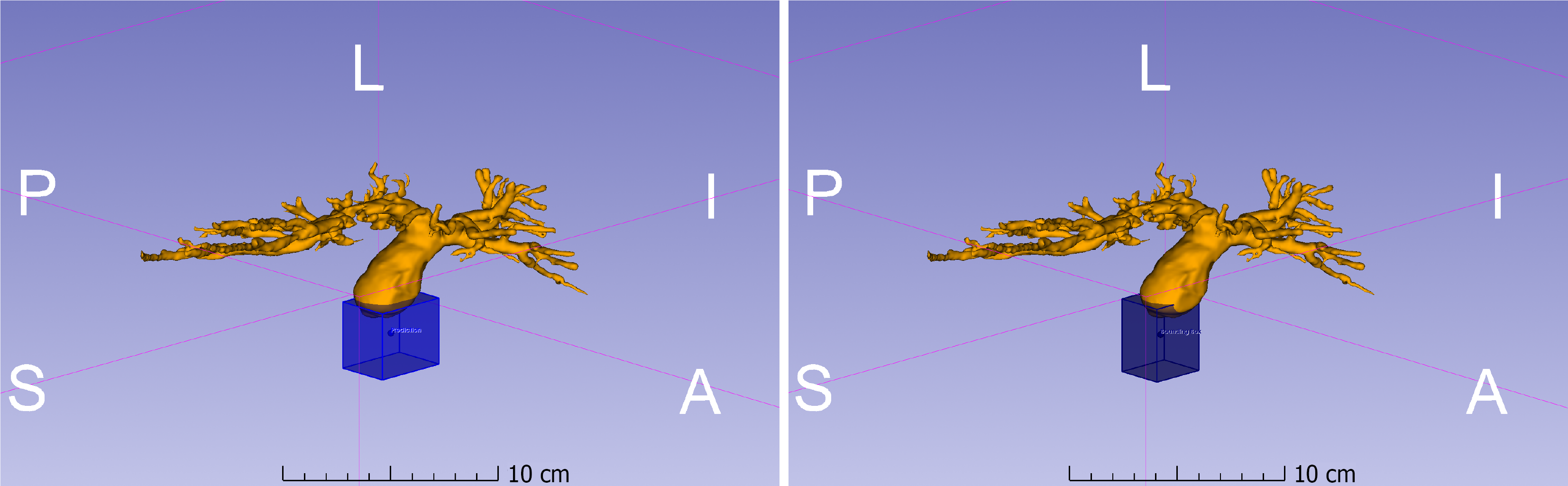
Figure 2 Three-dimensional stricture localization.
Manual annotation (left, blue) and MedYOLO prediction (right, dark blue) show that the predicted box covered the target ductal region but was broader than the manual label. Anatomical orientation is indicated, and a 10-cm scale bar is shown in each panel. A: Anterior; P: Posterior; L: Left; S: Superior; I: Inferior.
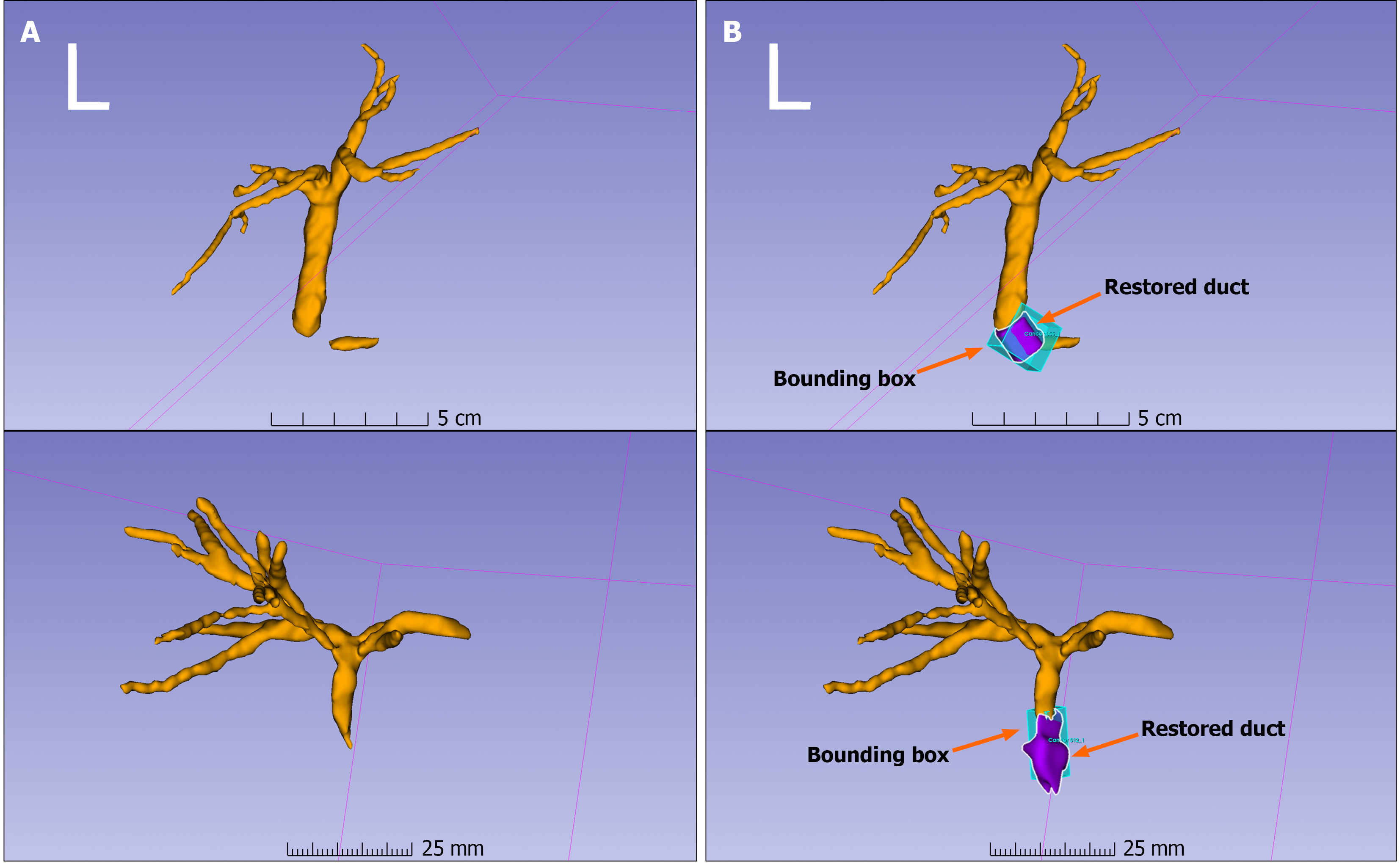
Figure 3 Representative morphology-restoration examples.
A: Original stenotic duct geometry; B: Restored target segment and localization box overlaid on the biliary tree. Arrows explicitly indicate the restored ductal segment and the corresponding ground-truth bounding-box region. The two rows show different representative examples.
- Citation: Chun JH, Hong JW, Hou JU, Lee KJ, Park DH, Cha HW, Park SW, Min SJ, Yu W. Artificial intelligence-driven three-dimensional modeling of biliary strictures using magnetic resonance cholangiopancreatography for quantitative assessment and stent planning. World J Gastroenterol 2027; 33(2): 125661
- URL: https://www.wjgnet.com/1007-9327/full/v33/i2/125661.htm
- DOI: https://dx.doi.org/10.3748/wjg.125661