
















Copyright: ©Author(s) 2026.
World J Gastroenterol. Dec 21, 2026; 32(47): 120094
Published online Dec 21, 2026. doi: 10.3748/wjg.v32.i47.120094
Published online Dec 21, 2026. doi: 10.3748/wjg.v32.i47.120094
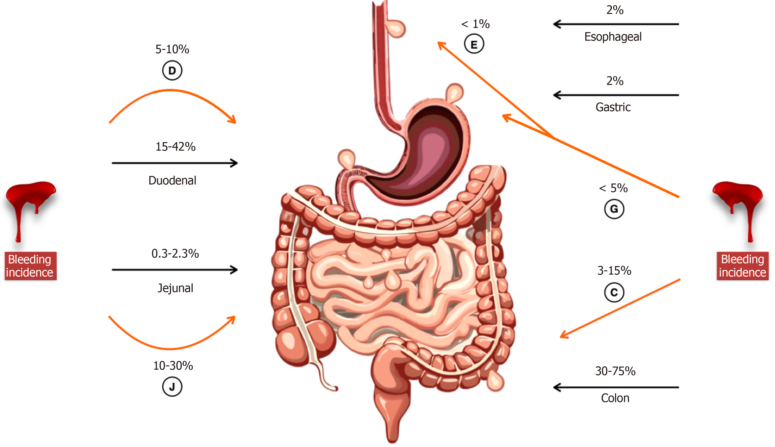
Figure 1 Anatomic distribution of diverticula and site-specific bleeding incidence.
Black arrows show gastrointestinal (GI) diverticula incidence. Incidence of diverticula bleeding is highlighted with a orange arrow. Jejunal diverticula accounts for a disproportionately higher bleeding rate (10%-30%). This distribution highlights the clinical relevance of jejunal diverticular disease in the assessment of unspecific GI bleeding. D: Duodenal; J: Jejunal; E: Esophageal; G: Gastric; C: Colon.
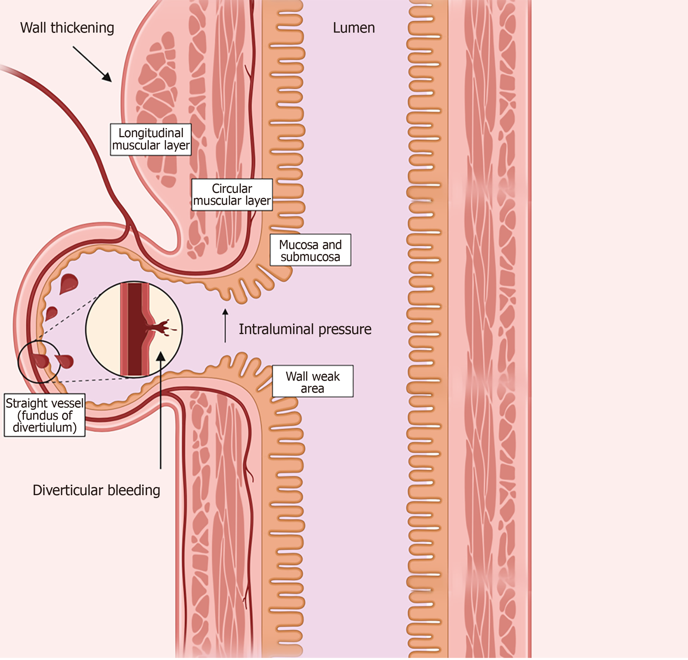
Figure 2 Development and bleeding of gastrointestinal diverticula.
Chronic intraluminal pressure at weak areas along with structural and functional alterations are key elements in gastrointestinal diverticula formation. Weak points are the areas where penetrating vessels traverse the circular muscle layer obliquely to stream within the submucosal plane. Structural alterations include abnormalities in collagen and elastin architecture, characterized by excessive collagen cross-linking, which leads to increased wall stiffness and diminished tensile integrity. Additionally, elastosis in the longitudinal muscular layer contributes to wall thickening. Bleeding occurs due to the erosion or rupture of the straight vessels (vasa recta).
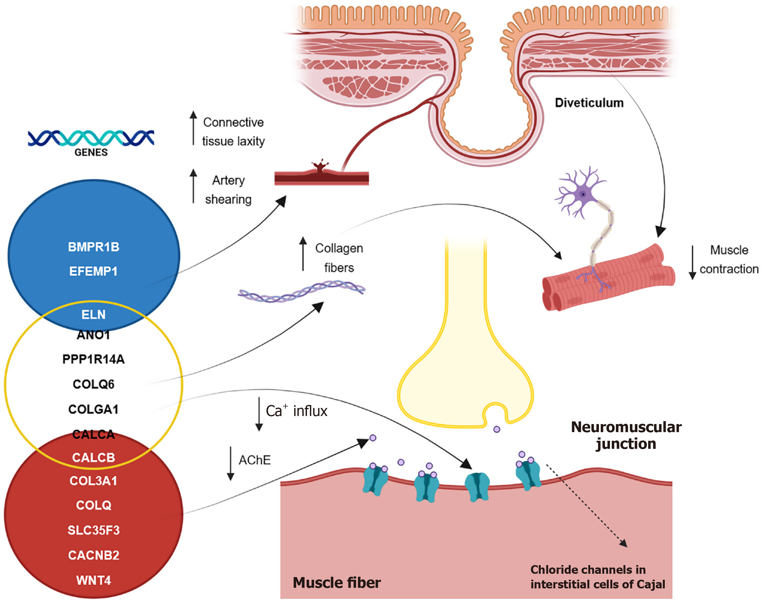
Figure 3 Biomolecular pathways involved in diverticular development.
Genes associated with connective tissue laxity including BMPR1B, EFEMP1 and ELN predispose to arterial shearing at the level of the diverticulum (blue circle). Genes involved in neuromuscular dysfunction such as ANO1, PPP1R14A, COLQ6, COL6A1, CALCA and CALCB (yellow circle) regulate calcium-activated chloride channels in the interstitial cells of Cajal, thereby affecting muscle contraction. Genes linked with vascular damage and structural integrity are CALCB, COL3A1, COLQ, SLC35F3, CACNB2 and WNT4. These genes contribute to vascular smooth muscle cell proliferation, increase collagen fibers between smooth muscle bundles and reduce AChE available at the neuromuscular junction leading to muscle weakness (red circle).
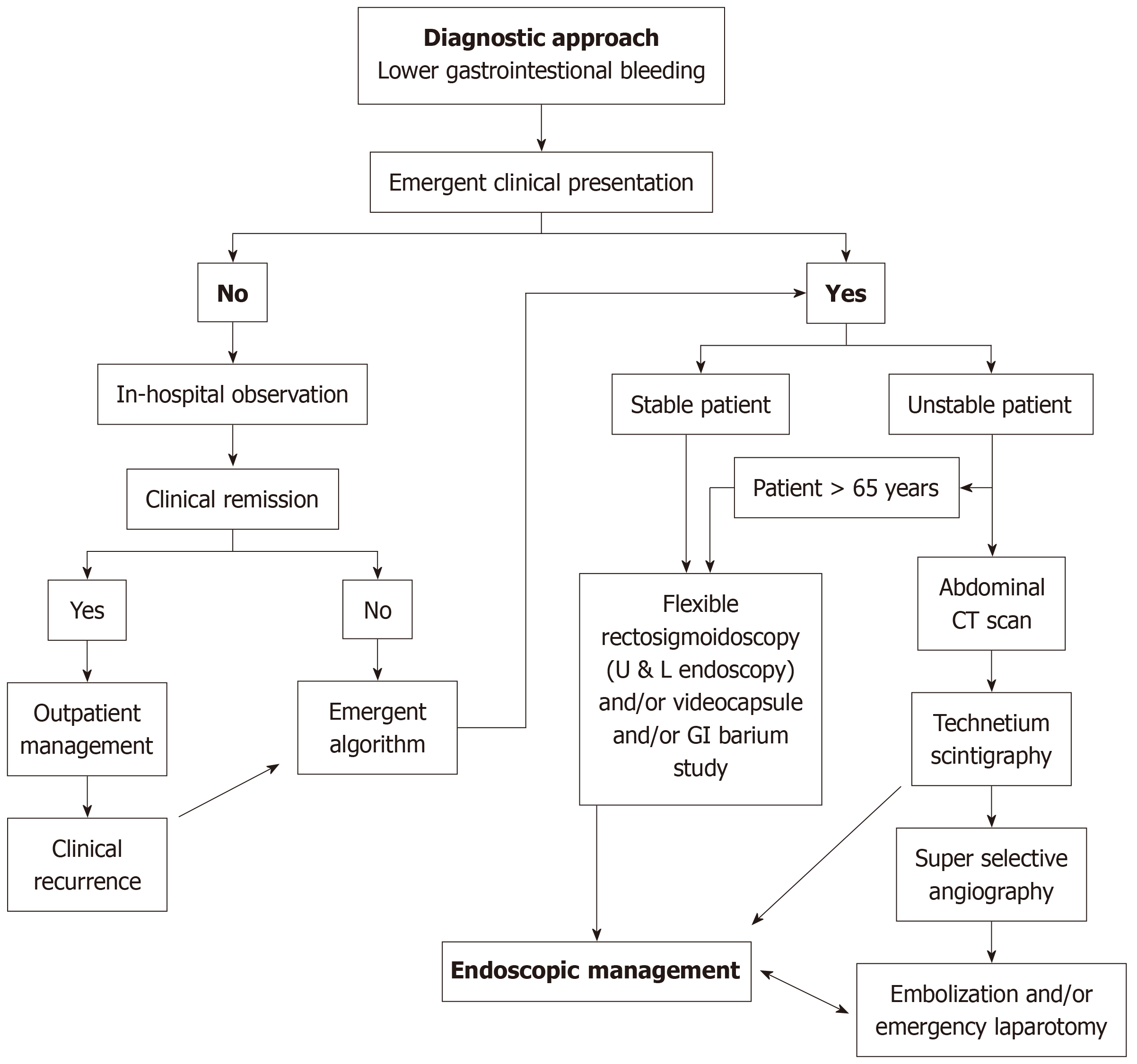
Figure 4 Diagnostic pathways for patients with suspected jejunal diverticular bleeding.
For patients without an emergent clinical presentation, in-hospital observation may be adequate. However, in cases of hemodynamic instability or ongoing hemorrhage, expeditious diagnostic assessment based on clinical urgency is imperative. When upper and lower endoscopies are negative, capsule endoscopy has a central role in the study of lower gastrointestinal bleeding. Once the diagnosis is established, endoscopic management should be pursued as the first-line therapeutic approach. U & L: Upper and lower; GI: Gastrointestinal; CT: Computed tomography.
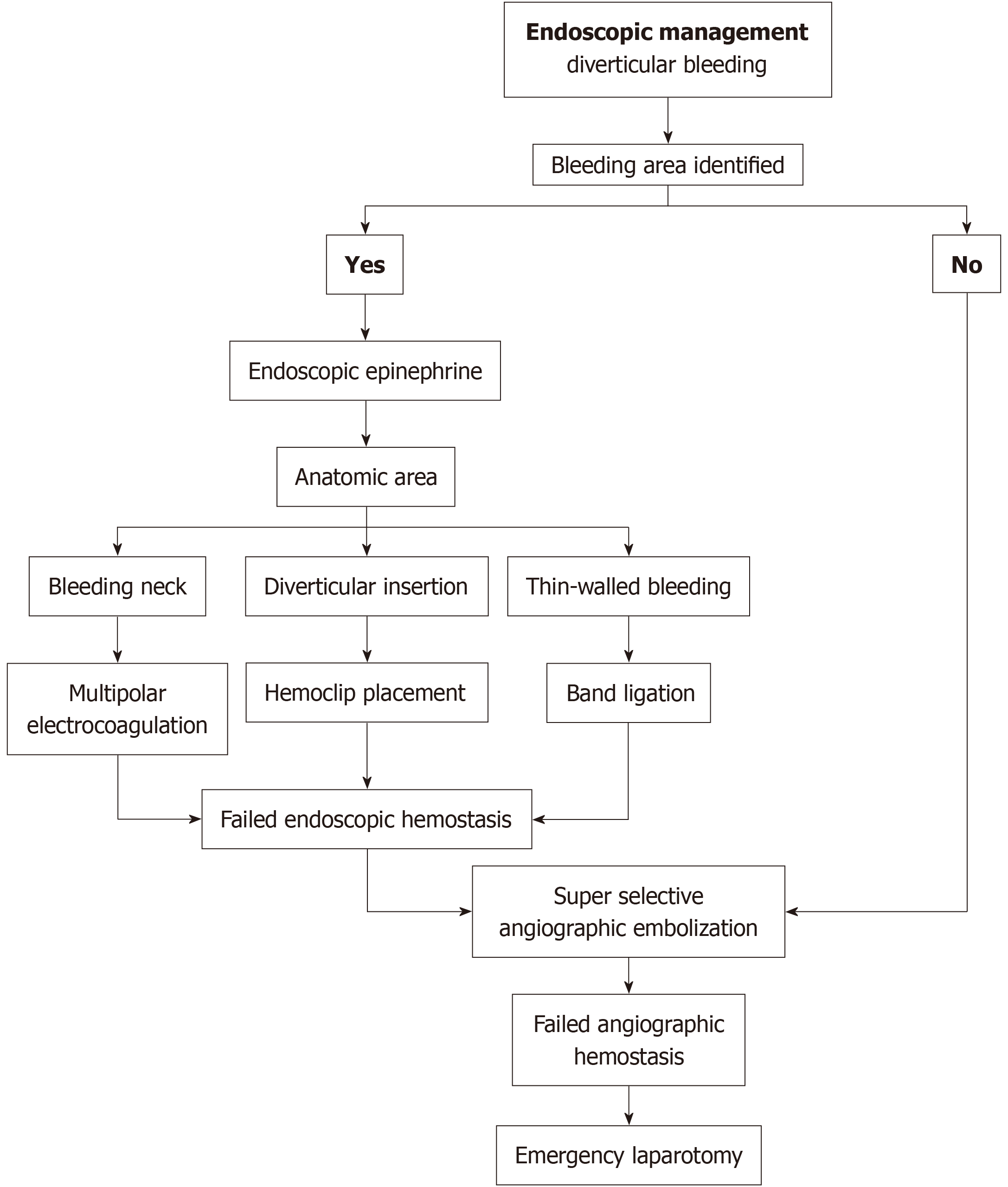
Figure 5 Endoscopic management algorithm for diverticular bleeding.
When the bleeding source is identified, appropriate endoscopic hemostatic modalities should be promptly implemented in accordance to the bleeding characteristics. In cases in which the bleeding site cannot be identified or endoscopic hemostasis fails, treatment escalation should not be delayed, and angiographic or surgical management must be considered based on clinical severity.
- Citation: Anaya-Prado R, Dominguez-Soto AV, Osorio-Medina KN, Ortiz-Vera RI, Gonzalez-Martinez D, Anaya-Fernández R, Portillo-Mendez R, Anaya-Fernández MM, Azcona-Ramirez CC, Guerrero-Palomera MA, Peña-Cuellar AJ, Soto-Hintze J, Reyna-Rodriguez B. Jejunal diverticula bleeding: A challenging diagnosis entity. World J Gastroenterol 2026; 32(47): 120094
- URL: https://www.wjgnet.com/1007-9327/full/v32/i47/120094.htm
- DOI: https://dx.doi.org/10.3748/wjg.v32.i47.120094