
















Copyright: ©Author(s) 2026.
World J Gastroenterol. Sep 7, 2026; 32(33): 118486
Published online Sep 7, 2026. doi: 10.3748/wjg.118486
Published online Sep 7, 2026. doi: 10.3748/wjg.118486
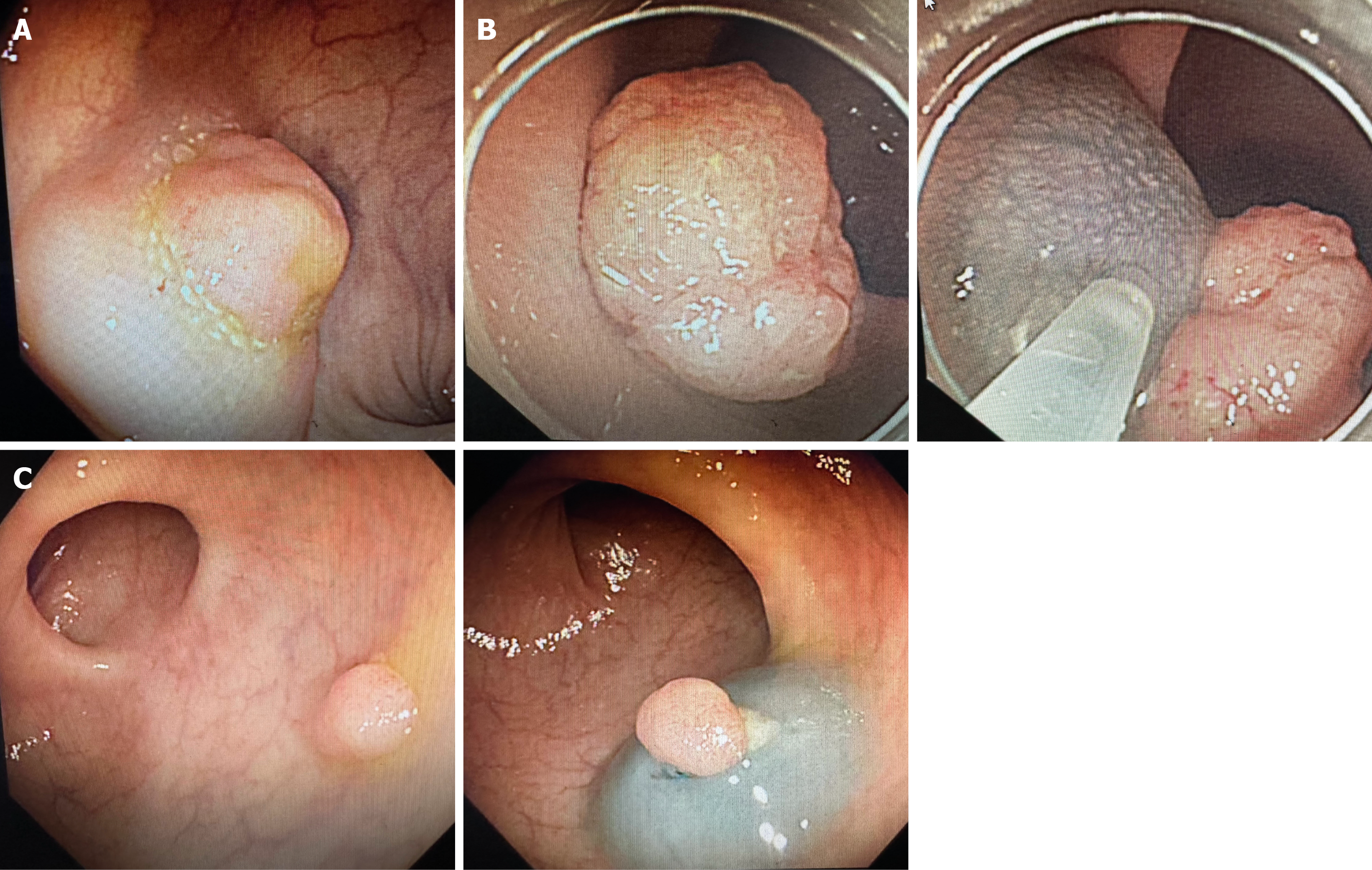
Figure 1 Endoscopic finding of chicken skin mucosa.
A: Adenoma with type 1 chicken skin mucosa (CSM) (confirmed before injection); B: Adenoma with type 2 CSM (confirmed after injection); C: Adenoma without CSM.
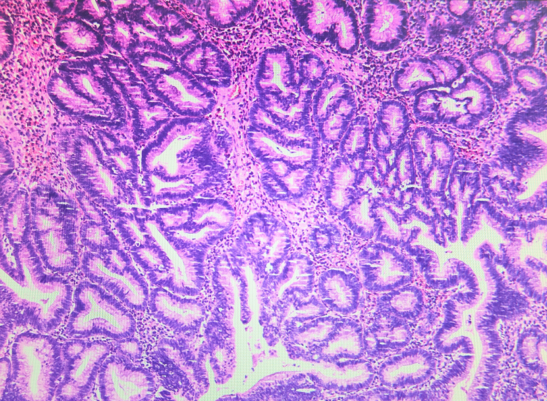
Figure 2 Histological characteristics of chicken skin mucosa, including lipid-filled macrophages in the lamina propria.
Hematoxylin and eosin, 40 ×.
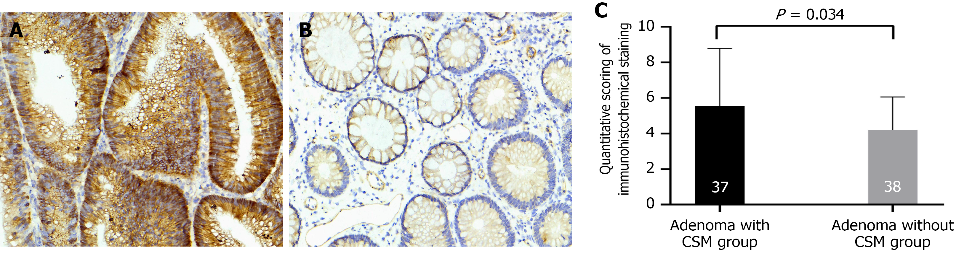
Figure 3 Expression of transmembrane member 16A protein by immunohistochemical staining.
A: Adenoma with chicken skin mucosa group; B: Adenoma without chicken skin mucosa group (original magnification, 400 ×); C: Quantitative scoring of immunohistochemical staining of transmembrane member 16A protein expression in each group (P = 0.034). CSM: Chicken skin mucosa.
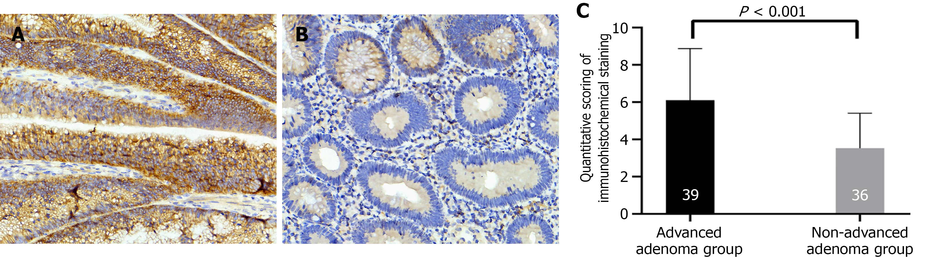
Figure 4 Expression of transmembrane member 16A protein by immunohistochemical staining.
A: Advanced adenoma group; B: Non-advanced adenoma group (original magnification, 400 ×); C: Quantitative scoring of immunohistochemical staining of transmembrane member 16A protein expression in each group (P < 0.0001).
- Citation: Wang RJ, Hu Y, Guo SB. Chicken skin mucosa is independently associated with advanced colorectal adenoma with transmembrane member 16A potentially involved in this association. World J Gastroenterol 2026; 32(33): 118486
- URL: https://www.wjgnet.com/1007-9327/full/v32/i33/118486.htm
- DOI: https://dx.doi.org/10.3748/wjg.118486