
















Copyright: ©Author(s) 2026.
World J Gastroenterol. Aug 14, 2026; 32(30): 119614
Published online Aug 14, 2026. doi: 10.3748/wjg.119614
Published online Aug 14, 2026. doi: 10.3748/wjg.119614
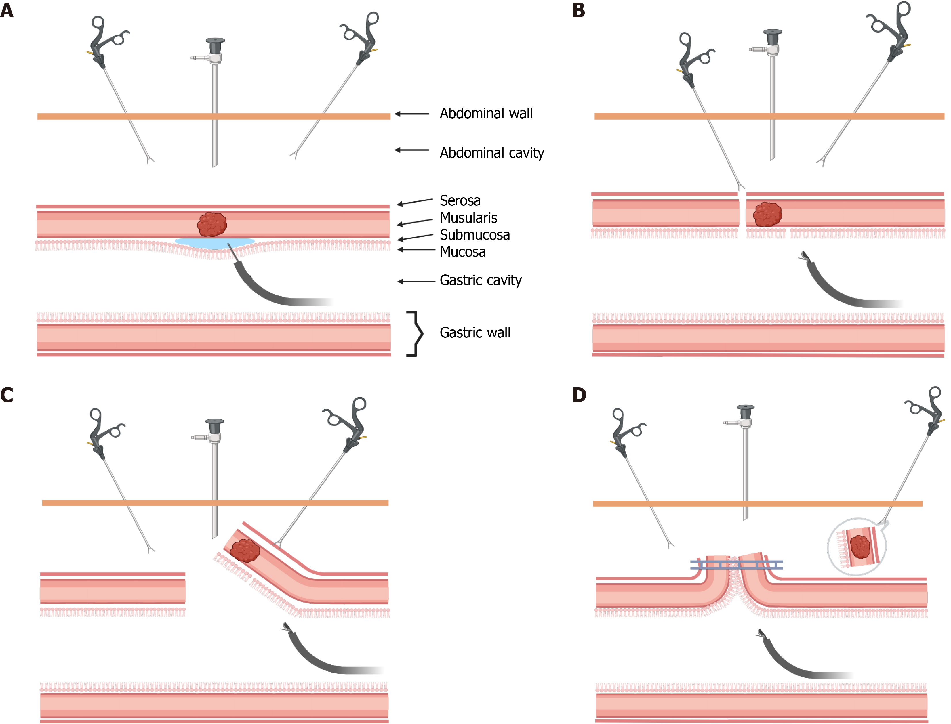
Figure 1 Classic laparoscopic endoscopic cooperative surgery operation steps.
A: Endoscopic injection of 10% glycerol into the submucosa; B: Endoscopic circumferential resection of the mucosa and submucosa, with extensive dissection of the seromuscular layer; C: Complete dissection of the remaining seromuscular layer under laparoscopy; D: Retrieval of the intact tumor tissue through a transabdominal approach, followed by closure of the incision using a laparoscopic mechanical stapler.
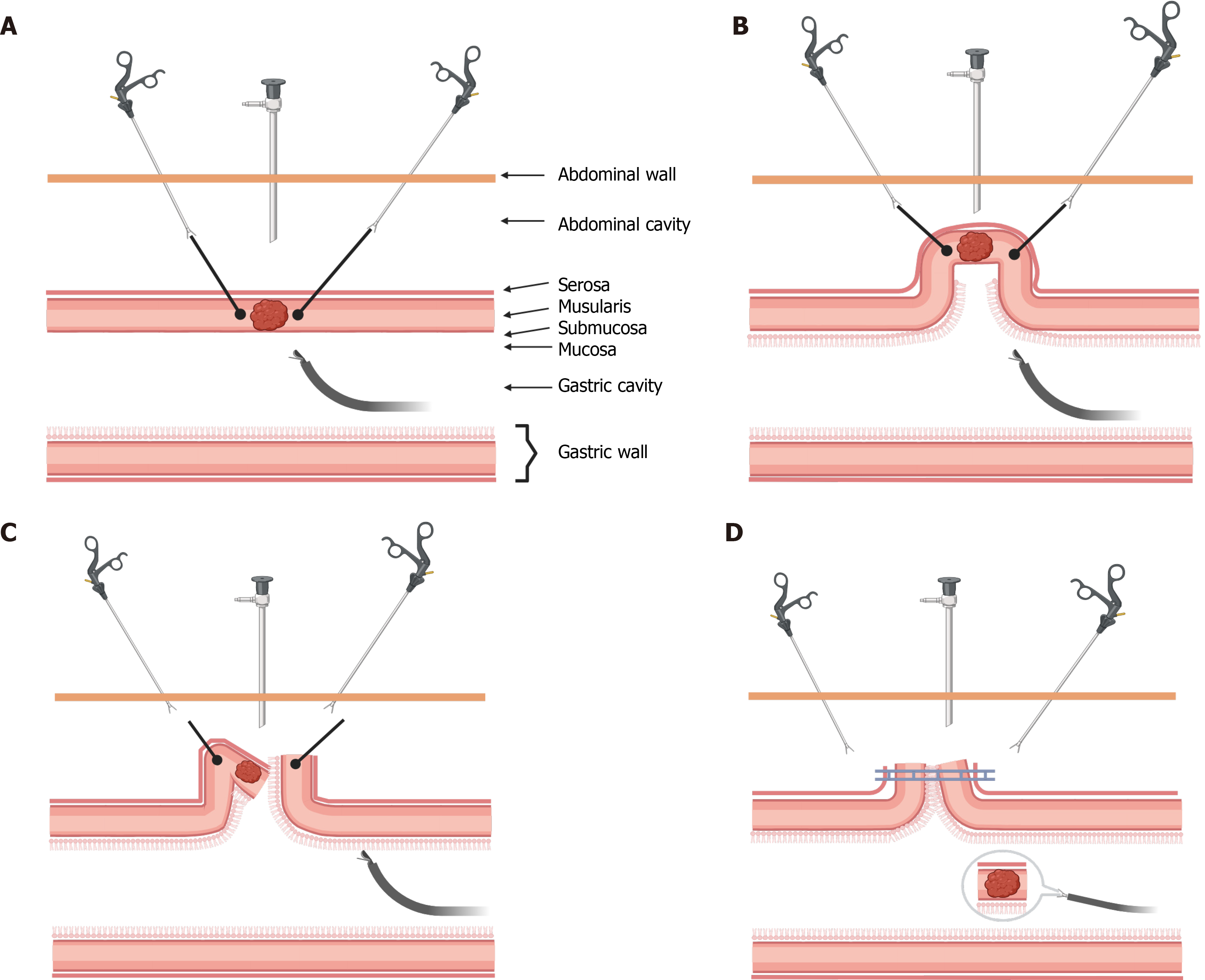
Figure 2 Inverted laparoscopic endoscopic cooperative surgery operation steps.
A: Endoscopic submucosal dissection and mucosal resection; B: Sutures suspend the gastric wall, forming a crown-like structure; C: Under laparoscopy, part of the seromuscular layer is dissected, and the tumor is inverted into the gastric cavity; D: Continuation of seromuscular layer dissection, followed by transoral extraction of the intact tumor tissue. The incision is then closed using a laparoscopic mechanical stapler.
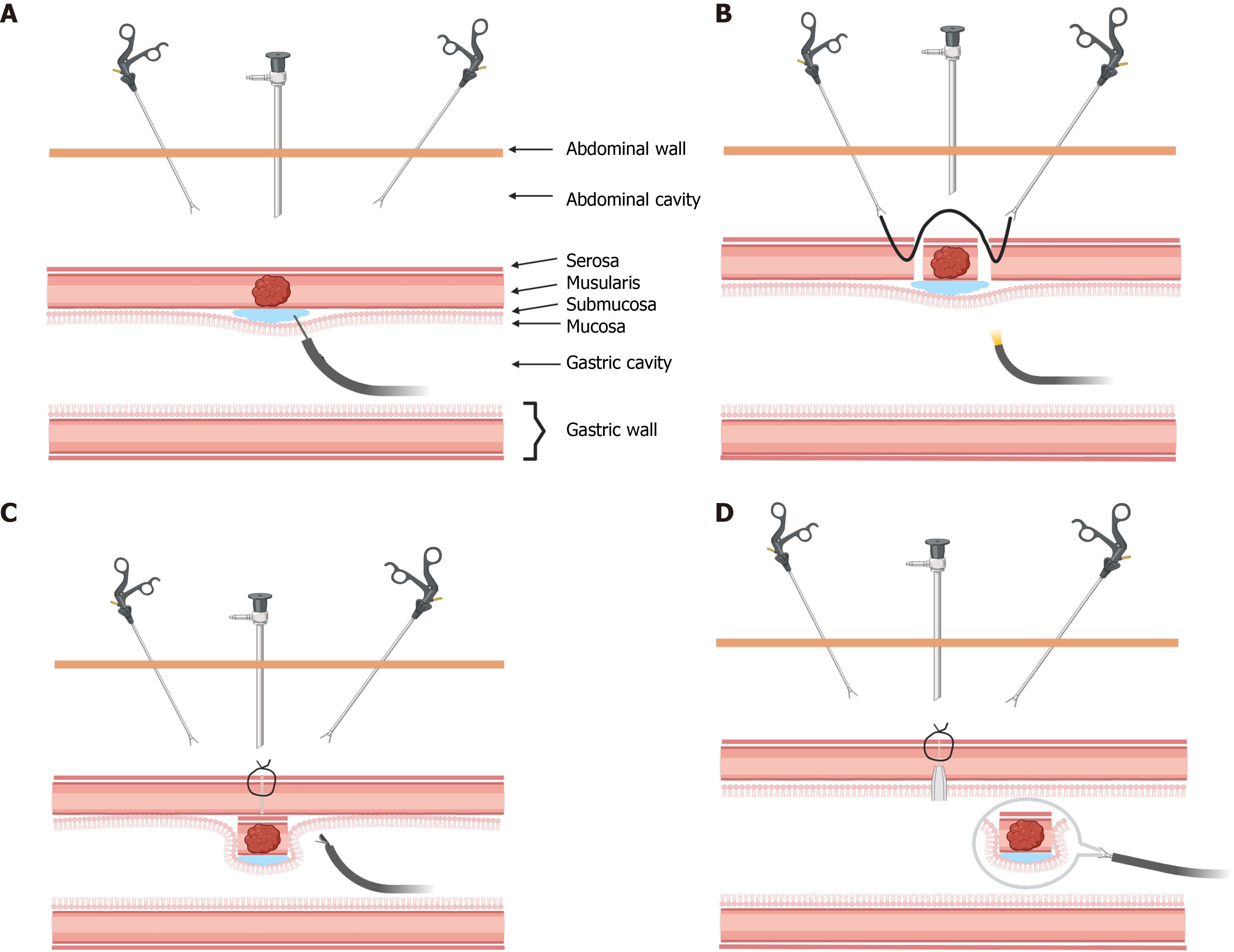
Figure 3 Non-exposed endoscopic wall inversion surgery operation steps.
A: Endoscopic injection of 10% glycerol into the submucosa; B: Circumferential incision of the seromuscular layer under laparoscopy, followed by linear suturing of the outer rim with 3-0 silk sutures; C: Tumor inversion into the gastric cavity; D: Complete removal of the tumor tissue through the oral cavity, with closure of the mucosal and submucosal layer wound edges using endoscopic forceps.
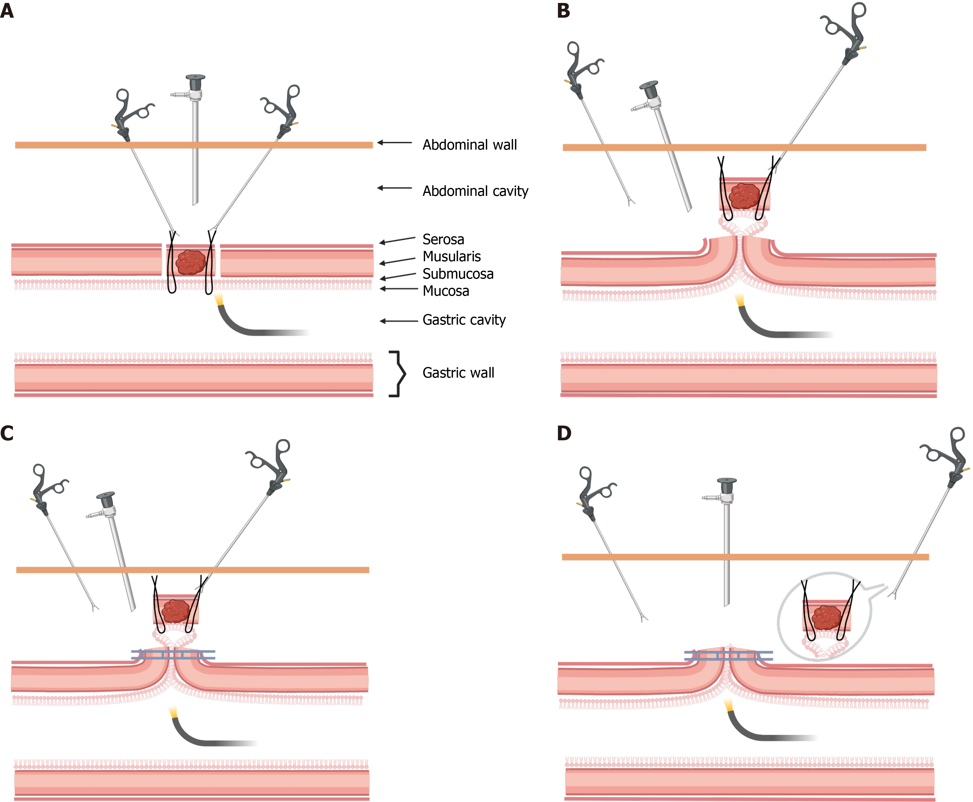
Figure 4 Combined laparoscopic and endoscopic approach for neoplasia with a non-exposure technique operation steps.
A: Placement of several 3-0 black silk sutures laparoscopically to fix the full thickness of the gastric wall, followed by dissection of the seromuscular layer along the outside of the sutures; B: The silk thread is pulled to create a natural “sealing area” in the tumor tissue; C: The mucosal tissue and free seromuscular layer are managed using a mechanical stapler; D: Complete removal of the tumor tissue through the abdominal cavity.
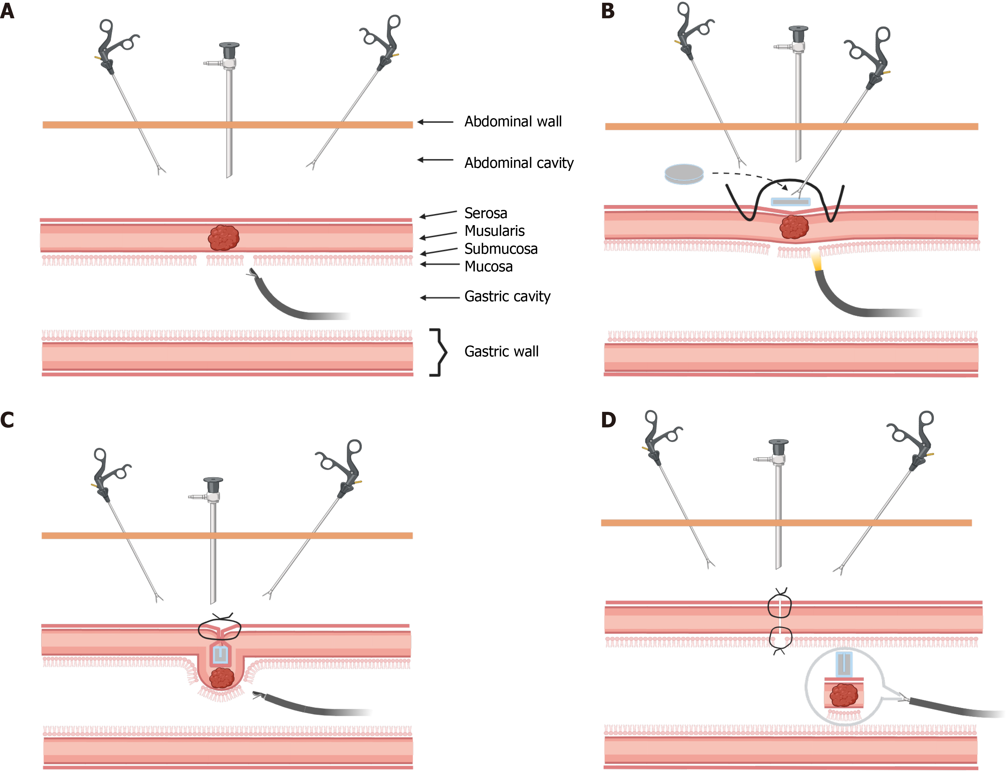
Figure 5 Closed laparoscopic endoscopic cooperative surgery operation steps.
A: Endoscopic circumferential resection of the mucosa and submucosa; B: Suturing of the seromuscular layers on both sides with 3-0 silk thread, fixing the gasket at the center of the suture line; C: The tumor tissue, along with the gasket, is inverted into the gastric cavity, and the seromuscular layer is circumferentially resected under endoscopic guidance; D: Complete removal of the tumor tissue and gasket through the mouth.
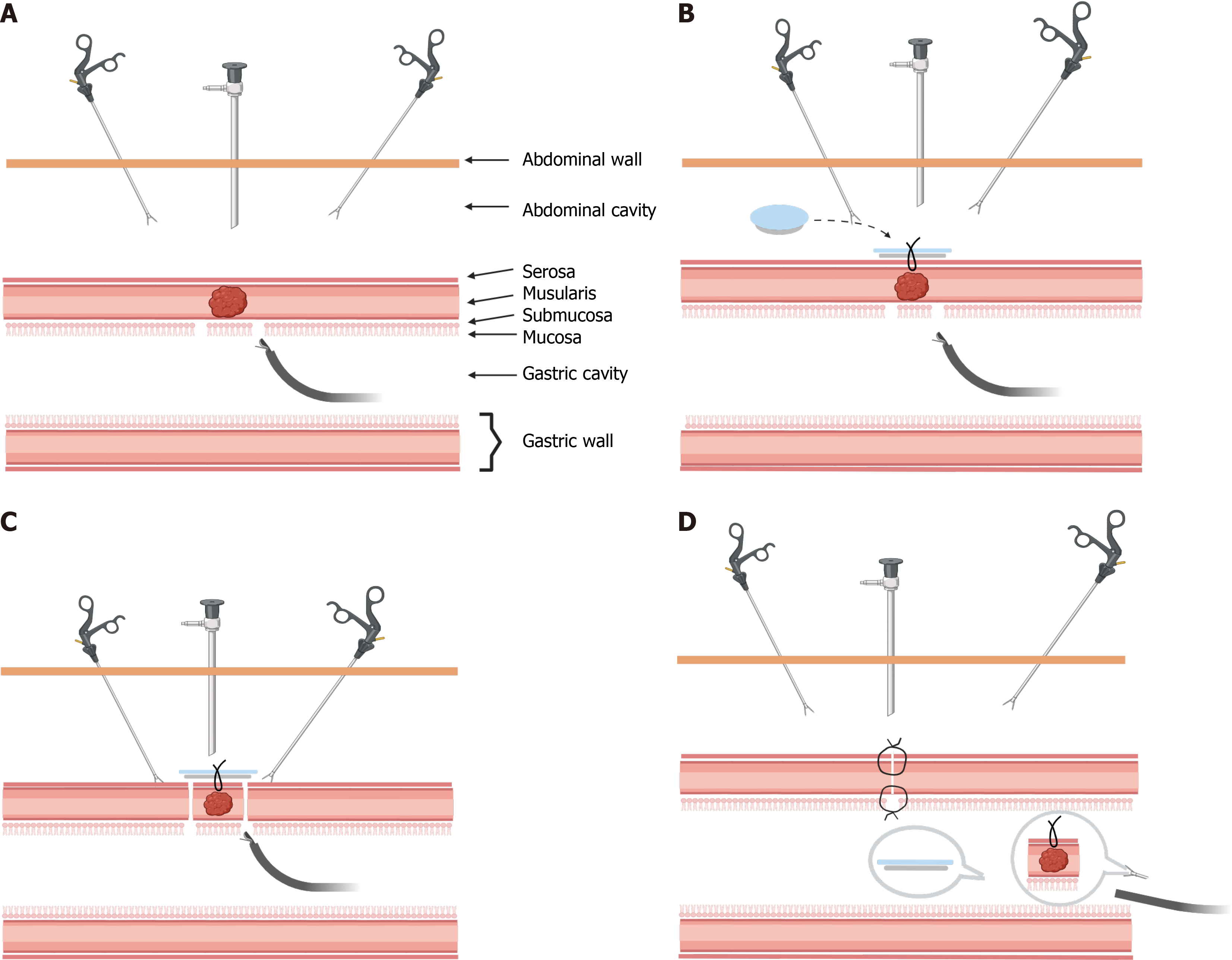
Figure 6 Sealed endoscopic full-thickness resection operation steps.
A: Endoscopic circumferential resection of the mucosa and submucosa; B: The silicone sheet is fixed to the serosa at the lesion site with 3-0 silk thread under laparoscopy; C: Endoscopic separation of the seromuscular layer; D: Complete removal of the tumor tissue, aided by silicone sheet spacers, through the mouth. The defect on the gastric wall is sutured linearly via laparoscopy.
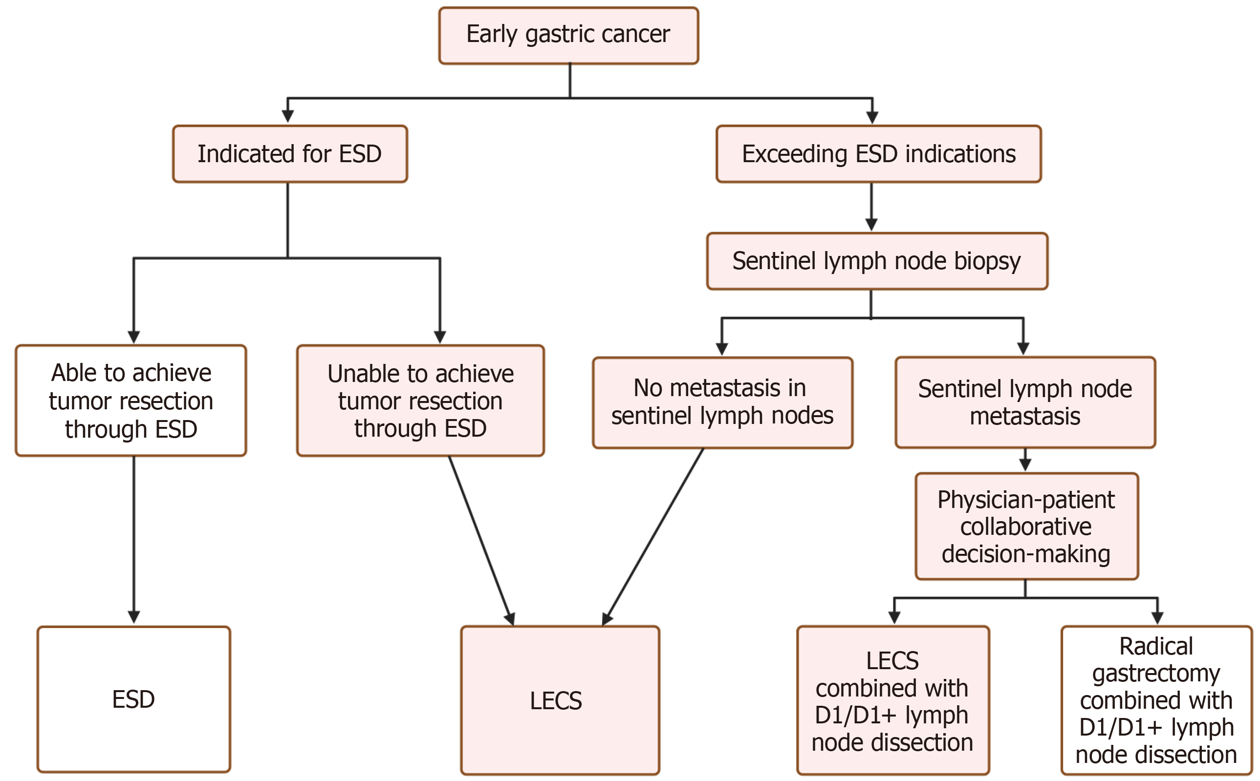
Figure 7 Laparoscopic endoscopic cooperative surgery combined with sentinel lymph node navigation surgery treatment for early gastric cancer surgical plan.
Red represents the laparoscopic endoscopic cooperative surgery technique; white represents other techniques. ESD: Endoscopic submucosal dissection; LECS: Laparoscopic endoscopic cooperative surgery; D1: First station; D1+: First station plus additional.
- Citation: Chen B, Han H, Zhang XX, Chen JX, Fan X. Research progress in laparoscopic endoscopic cooperative surgery for early gastric cancer: A narrative review. World J Gastroenterol 2026; 32(30): 119614
- URL: https://www.wjgnet.com/1007-9327/full/v32/i30/119614.htm
- DOI: https://dx.doi.org/10.3748/wjg.119614