
















Copyright: ©Author(s) 2026.
World J Gastroenterol. Jun 14, 2026; 32(22): 118810
Published online Jun 14, 2026. doi: 10.3748/wjg.v32.i22.118810
Published online Jun 14, 2026. doi: 10.3748/wjg.v32.i22.118810
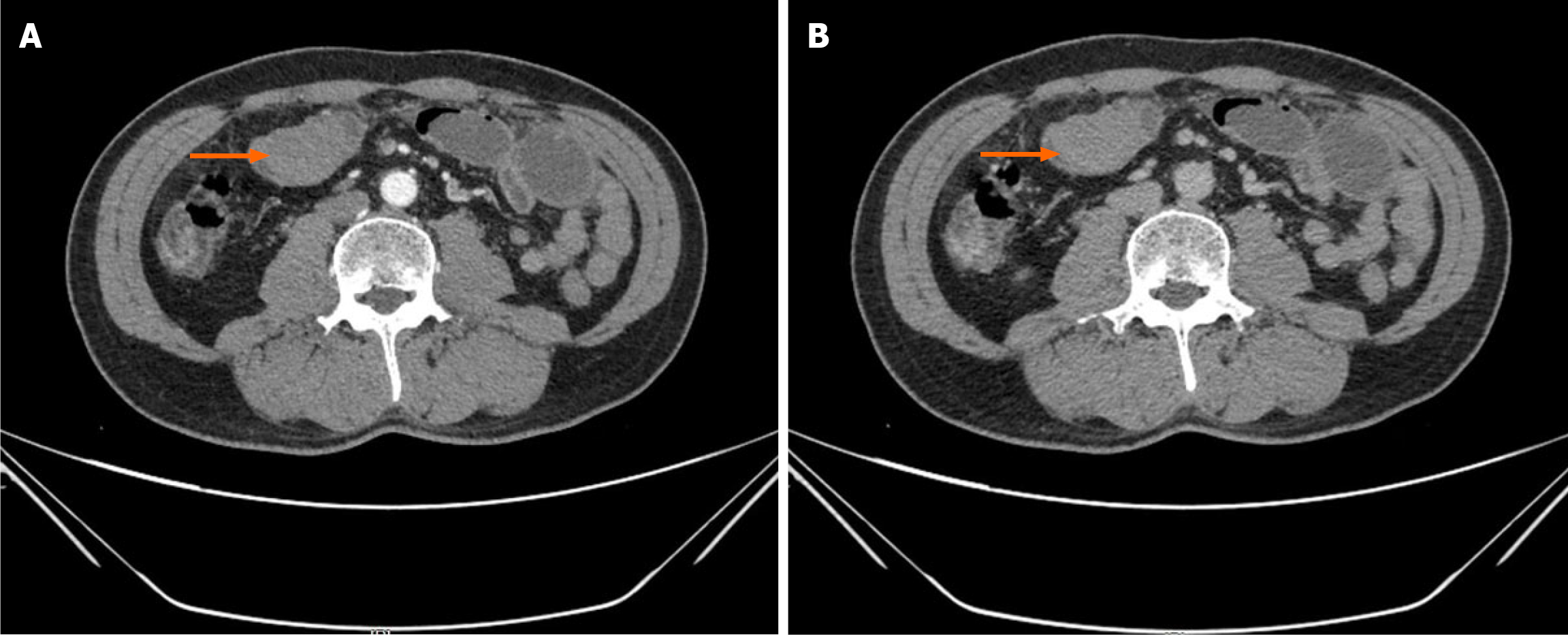
Figure 1 Contrast-enhanced abdominal computed tomography scan showing small intestinal mass.
A: Arterial phase axial image showing a well-defined enhancing mass (arrow) in the right mid-abdomen, causing proximal small bowel dilation; B: Portal venous phase confirms the soft-tissue density mass (arrow) with associated small bowel obstruction, initially suspected to be a gastrointestinal stromal tumor.
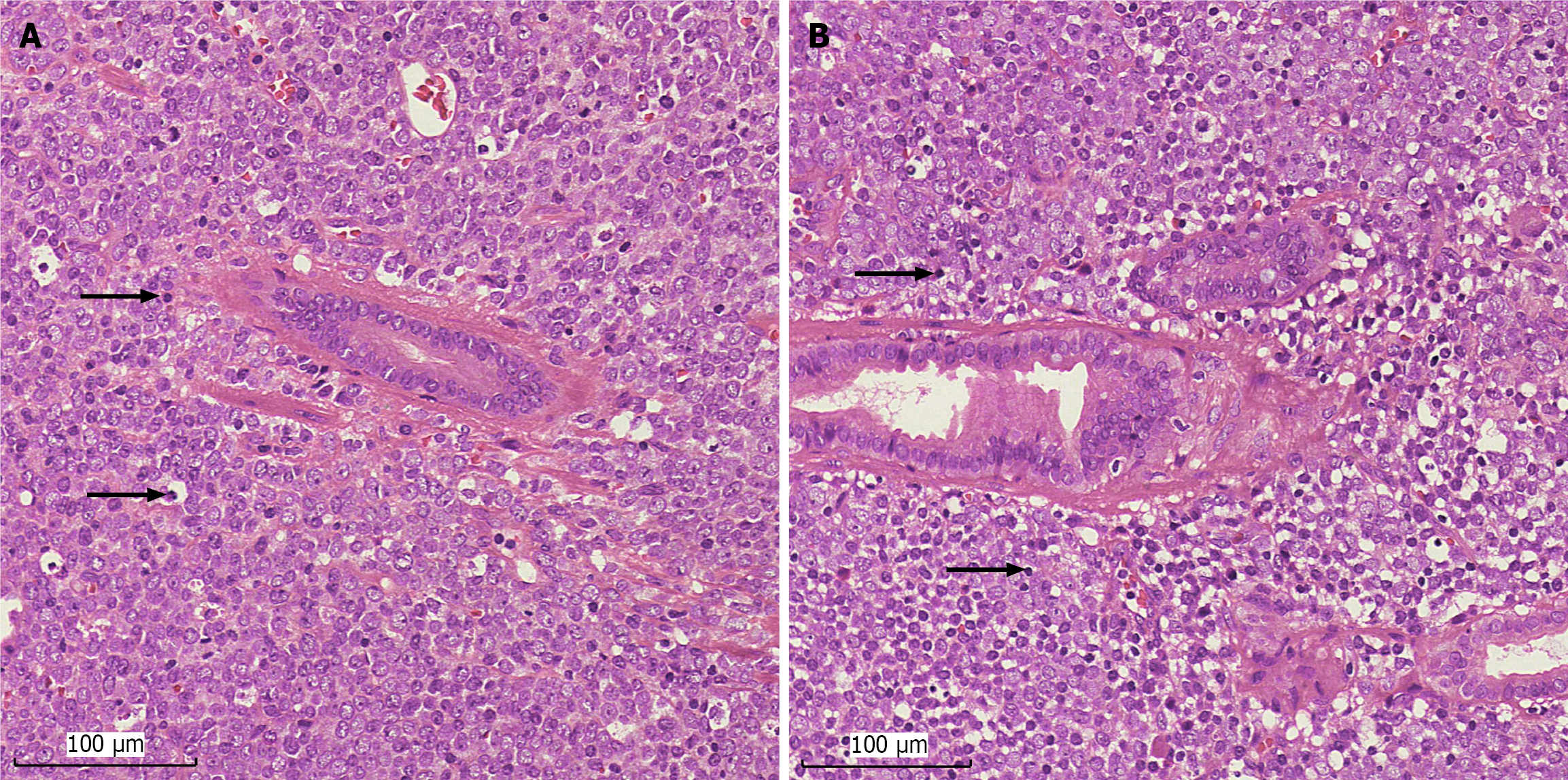
Figure 2 Histopathology of (hematoxylin and eosin staining) the resected small intestinal mass.
A and B: Histopathology of the resected small intestinal mass (× 200; scale bar, 100 μm). Tumor cells of medium size with a fairly uniform morphology infiltrate and grow diffusely in the intestinal wall (arrows). They have large, round or ovoid nuclei and scant cytoplasm. Nuclear chromatin is finely and evenly distributed, and in some cases, nucleoli of varying sizes are evident.
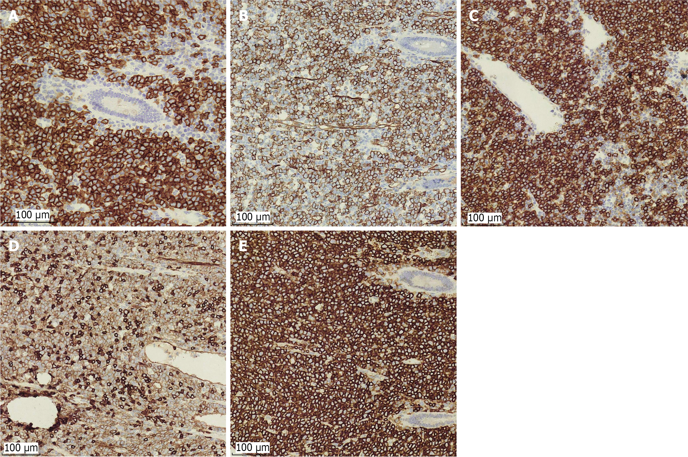
Figure 3 Immunohistochemical staining of tumor cells.
A: Myeloperoxidase strong cytoplasmic positivity confirming myeloid lineage; B-E: The tumor shows positive immune responses to myeloid markers cluster of differentiation (CD) 34 (B), CD117 (C), CD123 (D), and CD43 (E) (× 200; scale bar, 100 μm).
- Citation: Lin JQ, Liu HZ, Li ZW, Yang K. Intestinal myeloid sarcoma mimicking gastrointestinal stromal tumor without leukemic manifestations: A case report and review of literature. World J Gastroenterol 2026; 32(22): 118810
- URL: https://www.wjgnet.com/1007-9327/full/v32/i22/118810.htm
- DOI: https://dx.doi.org/10.3748/wjg.v32.i22.118810