©The Author(s) 2025.
World J Gastroenterol. Nov 7, 2025; 31(41): 112794
Published online Nov 7, 2025. doi: 10.3748/wjg.v31.i41.112794
Published online Nov 7, 2025. doi: 10.3748/wjg.v31.i41.112794
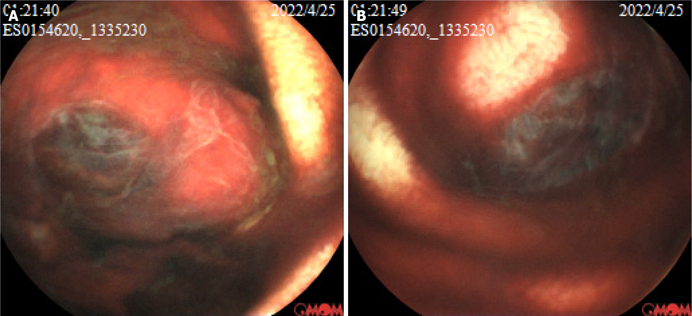
Figure 1 The capsule endoscopy examination shows a protruding lesion with a diameter of approximately 1.
5 cm, ulcerations, and dark red bloodstains in the jejunum. A and B: The morphological manifestations of the lesion from different perspectives.
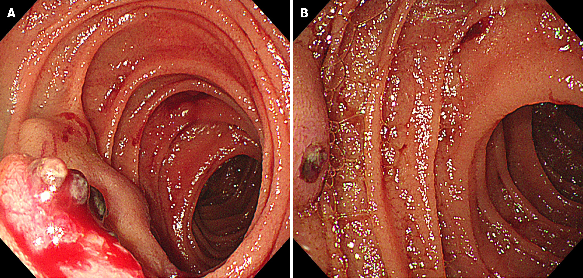
Figure 2 The oral single-balloon enteroscopy reveals two adjacent protruding lesions with surface erosion, blood clots, and a small amount of blood.
A: The surface morphology of the labial lesion and the local morphology of the anal lesion; B: The surface morphology of the anal lesion.
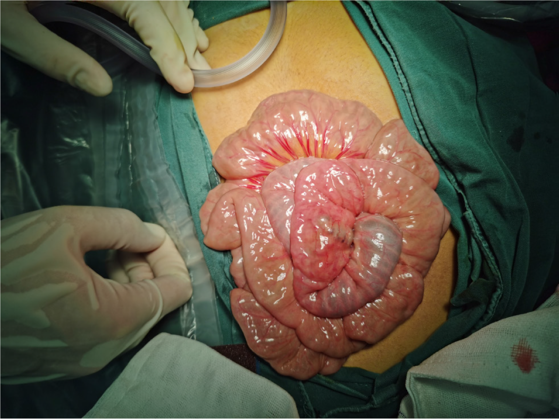
Figure 3
Intraoperatively, the serosal surface of the intestine was smooth, and the local nodular shadows with some parts of the intestines appeared blackish purple, suggestive of blood within the intestinal lumen.
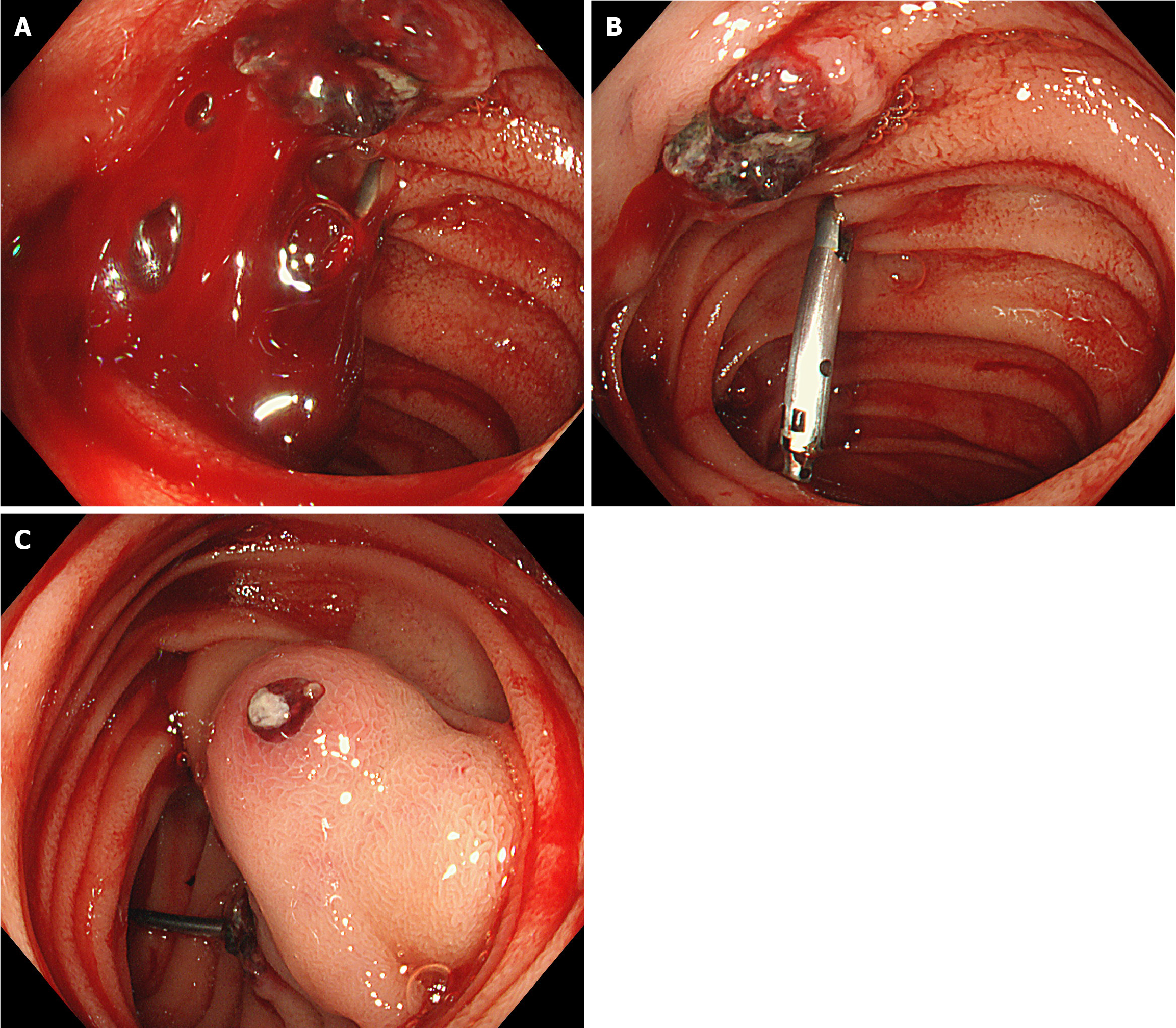
Figure 4 The manifestations of small intestinal lesions under intraoperative enteroscopy.
A: The intraoperative enteroscopy through the incision of the intestinal segment on the lesion’s anal side reveals numerous fresh red bloodstains on the lesion’s surface; B: After rinsing, the local ulceration, active bleeding, and titanium clip on the oral side of the lesion were visible; C: On the anal side, which had a submucosal protruding mass, surface ulceration, adhering dark red bloodstains, and white coating were visible.
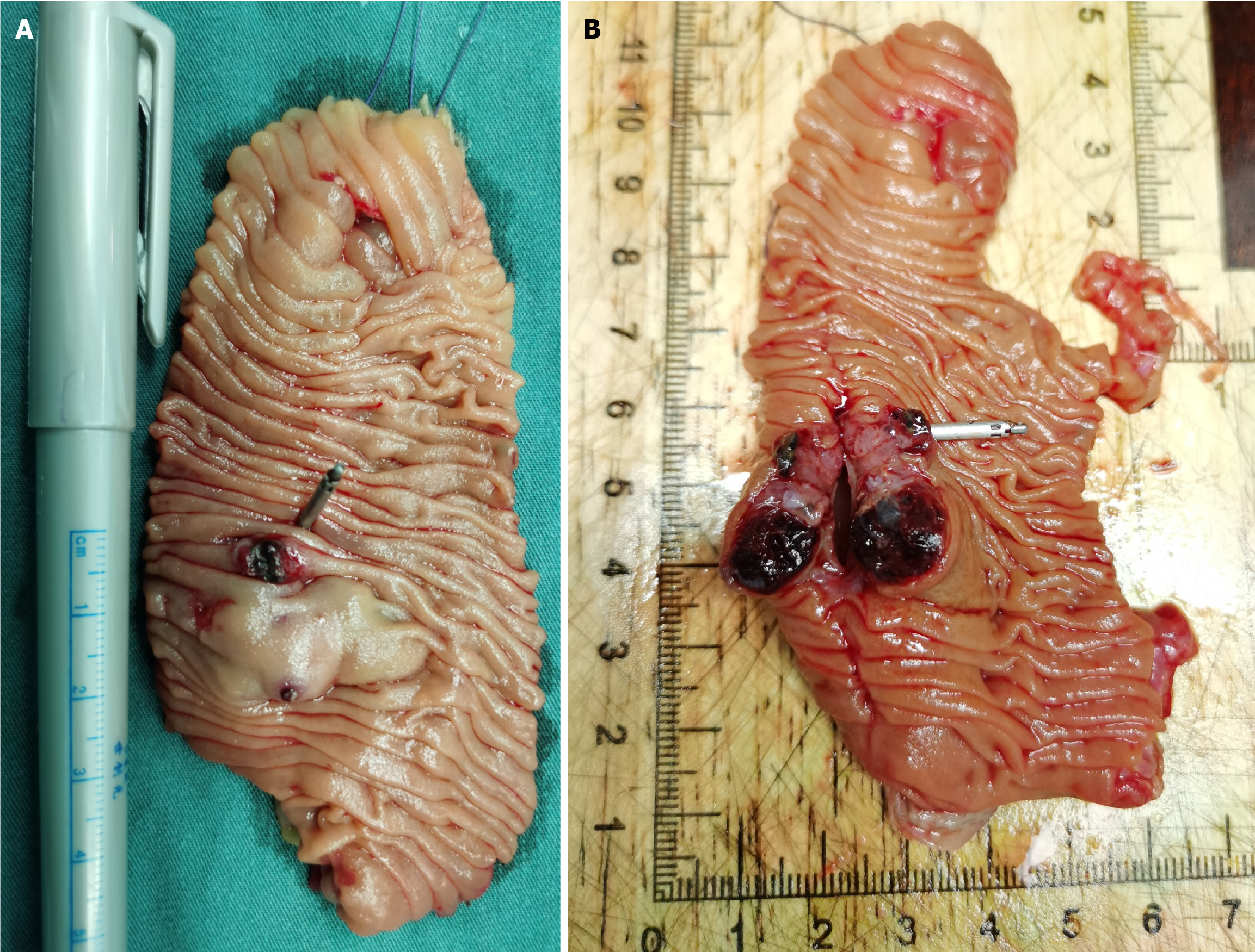
Figure 5 Postoperative gross specimen.
A: Upon opening the resected jejunum, a submucosal protruding mass with two ulcerations of different sizes was visible; B: After the lesion was incised, the tumor was observed to be grayish-white and grayish-red, with purple-red blood clots.
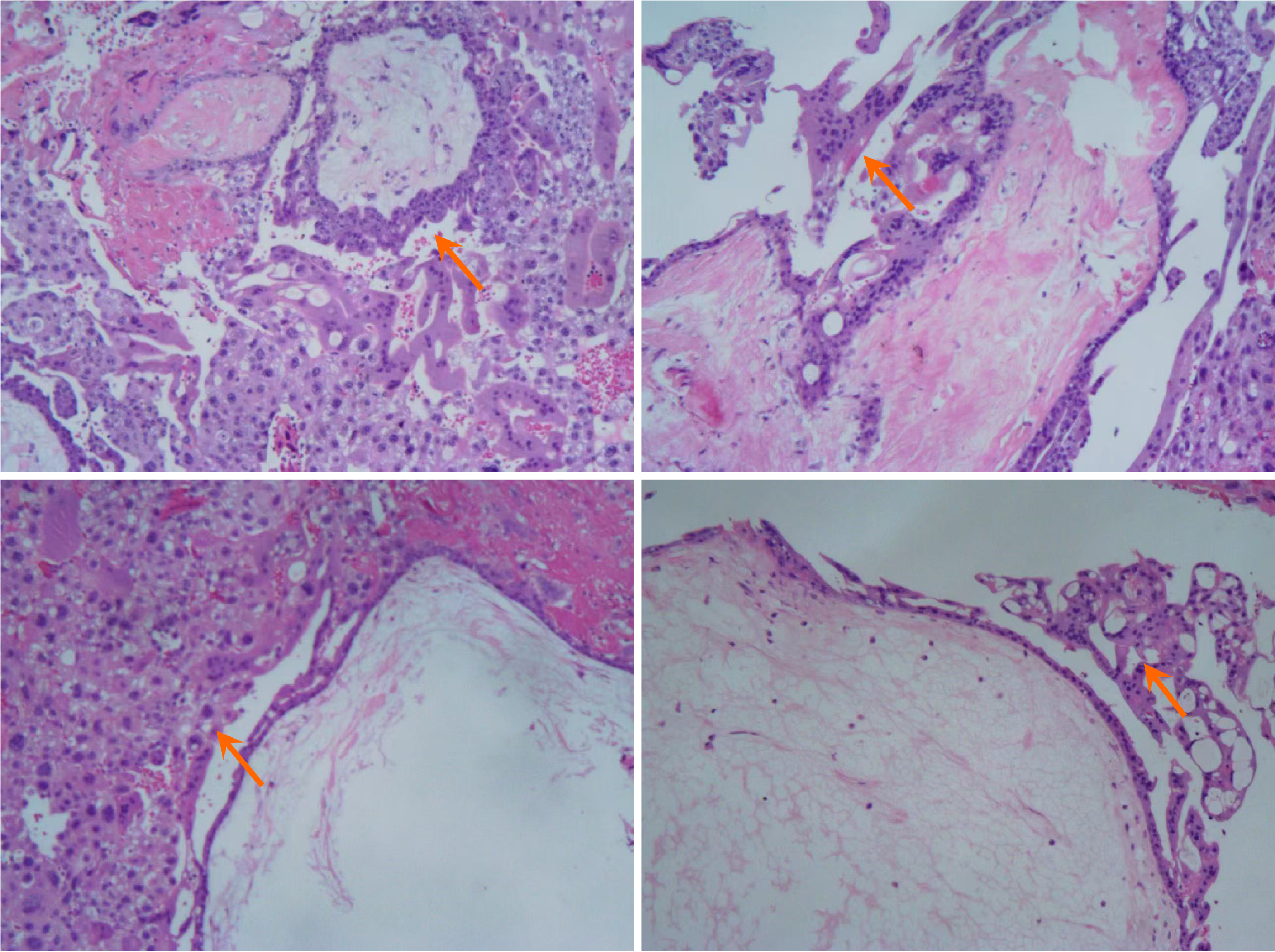
Figure 6 On the histopathology of the resected specimen, the hemorrhage, necrosis, and hyperplasia of trophoblastic cells were ob
- Citation: Yu WH, Zong Y, Zhou JW, Xu GQ. Uncommon causes of acute small intestinal bleeding-invasive mole: A case report and review of literature. World J Gastroenterol 2025; 31(41): 112794
- URL: https://www.wjgnet.com/1007-9327/full/v31/i41/112794.htm
- DOI: https://dx.doi.org/10.3748/wjg.v31.i41.112794


