©The Author(s) 2025.
World J Gastroenterol. Sep 14, 2025; 31(34): 110611
Published online Sep 14, 2025. doi: 10.3748/wjg.v31.i34.110611
Published online Sep 14, 2025. doi: 10.3748/wjg.v31.i34.110611
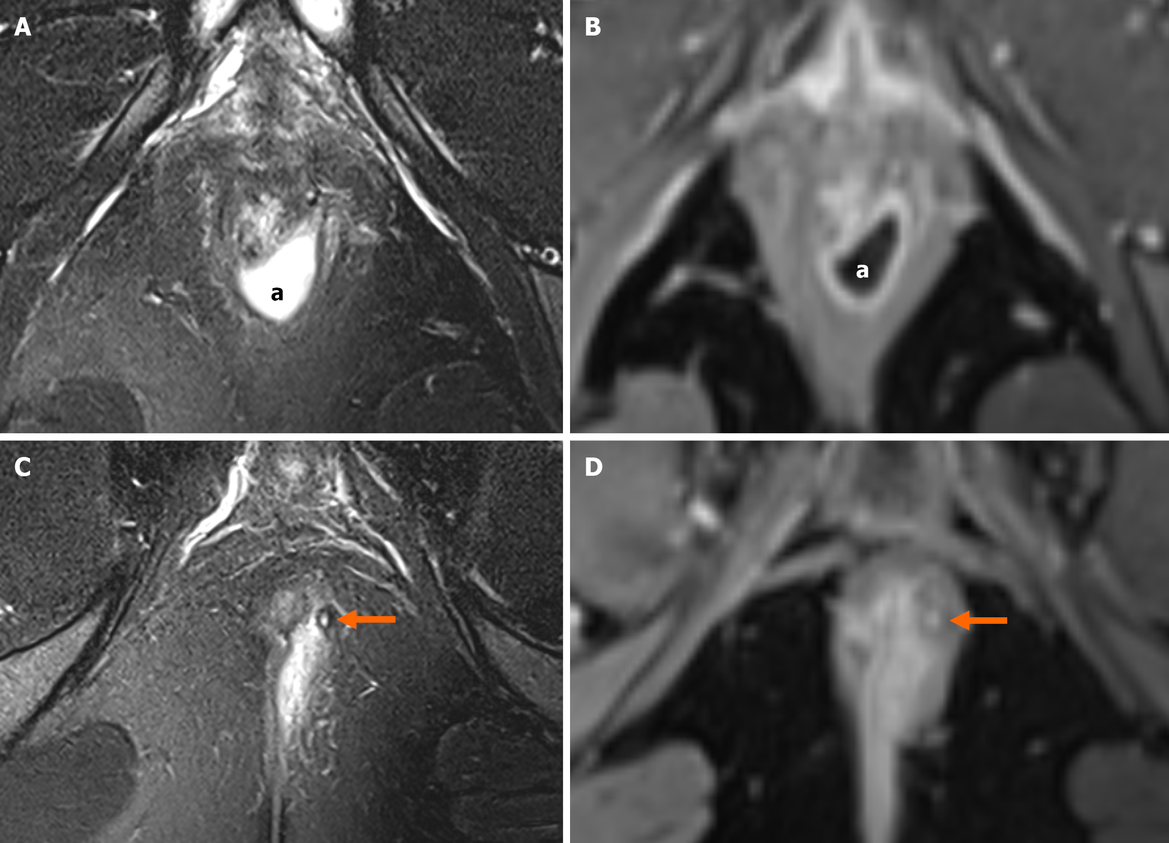
Figure 1 Magnetic resonance imaging of the pelvis demonstrating an intersphincteric fistula with an associated collection.
A: Axial T2 fat-saturated image: Intersphincteric collection (“a” sign) with T2 hyperintense content; B: Axial T1 fat-saturated post-gadolinium image: This collection demonstrates rim enhancement surrounding a central low-signal area, consistent with pus; C: Axial T2 fat-saturated image: Intersphincteric tract (arrow) demonstrates a central hyperintense signal with a hypointense rim; D: Axial T1 fat-saturated post-gadolinium image: The tract contents show enhancement without rim enhancement, consistent with granulation tissue within the tract and a fibrous tract wall.
Figure 2 Magnetic resonance imaging of the pelvis demonstrating a posterior branching fistula.
A: Axial T2-weighted fat-saturated; B: Coronal T2-weighted fat-saturate; C: Axial T1-weighted fat-saturated post-contrast T1-weighted. Posterior branching fistula with internal opening posteriorly at 6 o’clock. A fibrotic right-sided branch is noted at 7 o’clock demonstrating low T2 signal of the tract (A and B, arrows) and no contrast enhancement (C, arrow). An inflamed left-sided branch is noted at 5 o’clock demonstrating high T2 signal within the tract (A and B, arrowheads) and pronounced rim enhancement with central low signal related to fluid/pus (C, arrowhead).
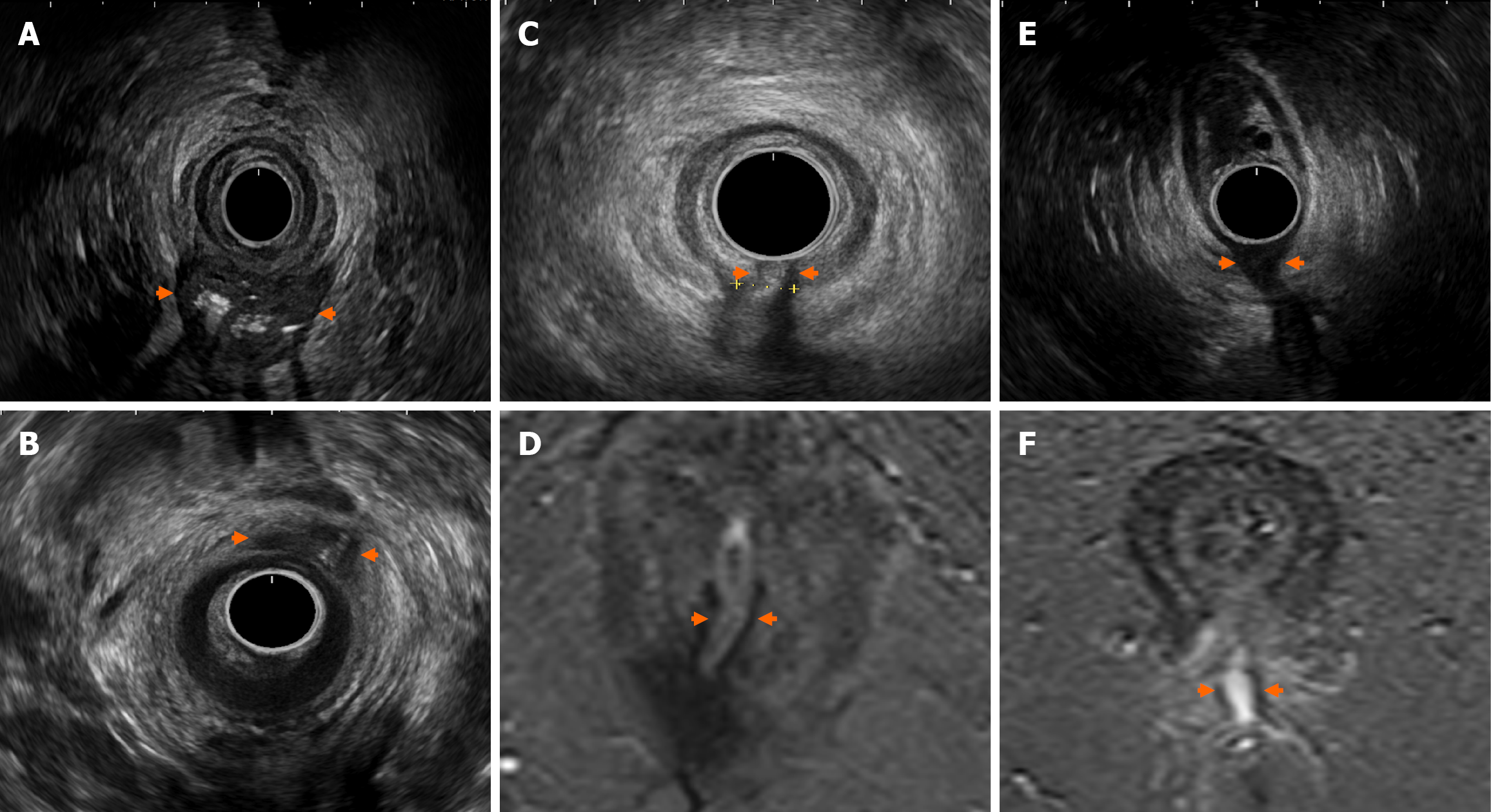
Figure 3 Evaluating perianal fistulising Crohn’s disease using endoanal ultrasound.
A: Large intersphincteric abscess with gas content (reverberation artefact from gas) posterior; B: Anterior intersphincteric abscess; C and D: Posterior midline transphincteric fistula with corresponding magnetic resonance imaging; E and F: Low transphincteric fistula with corresponding magnetic resonance imaging. Arrows delineate perianal pathology seen on endoanal ultrasound.
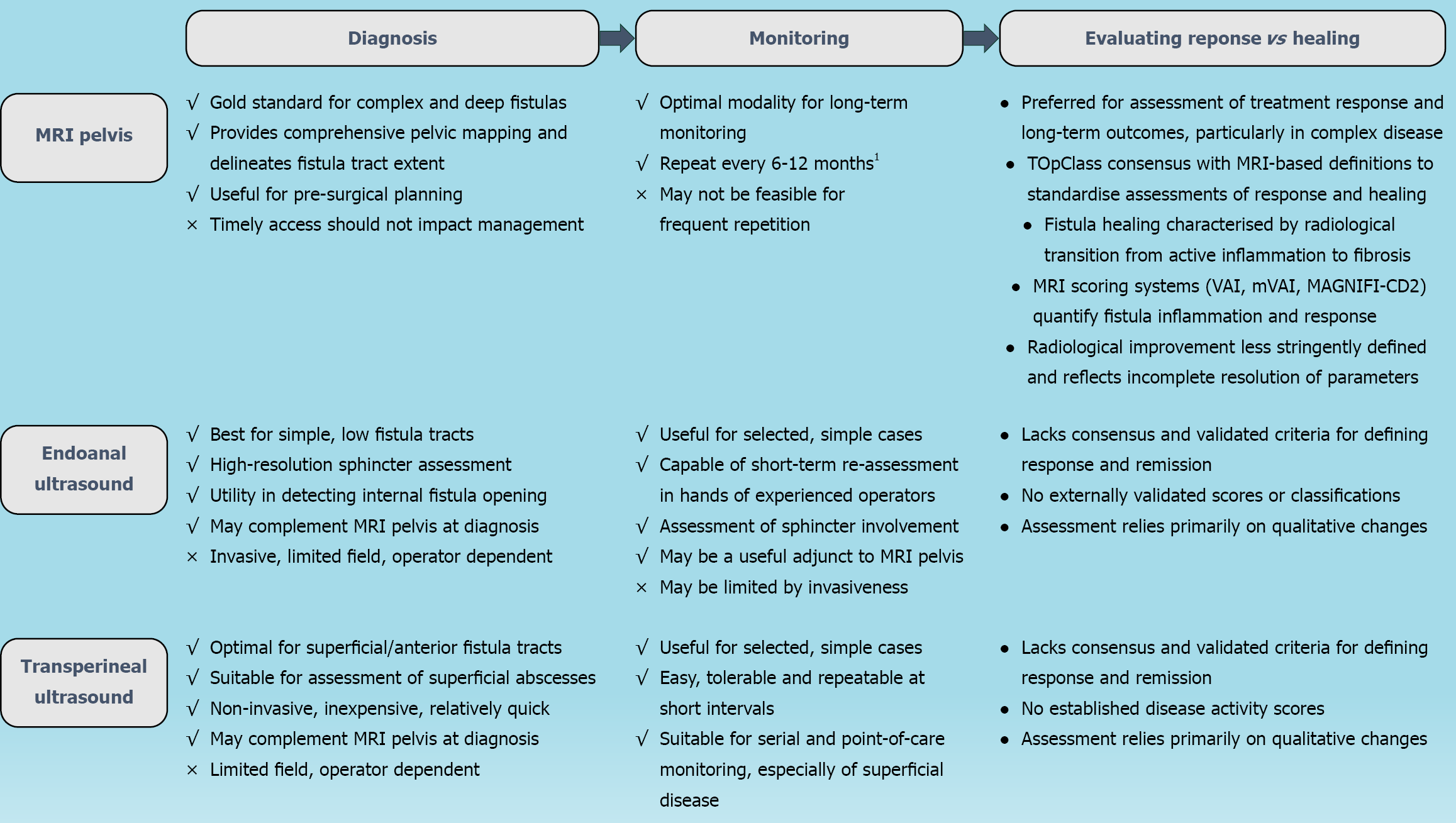
Figure 4 Imaging in perianal fistulising Crohn’s disease.
MRI: Magnetic resonance imaging; TOpClass: Treatment Optimisation and CLASSification; VAI: Van Assche Index; mVAI: Modified Van Assche Index; MAGNIFI-CD: Magnetic resonance imaging-based index for Crohn’s disease. 1Sooner if persistent symptoms, or planned treatment change; 2Supported by TOpClass.
- Citation: Habeeb H, Chen L, De Kock I, Bhatnagar G, Kutaiba N, Vasudevan A, Srinivasan AR. Imaging in perianal fistulising Crohn’s disease: A practical guide for the gastroenterologist. World J Gastroenterol 2025; 31(34): 110611
- URL: https://www.wjgnet.com/1007-9327/full/v31/i34/110611.htm
- DOI: https://dx.doi.org/10.3748/wjg.v31.i34.110611


