Copyright
©The Author(s) 2025.
World J Gastroenterol. May 28, 2025; 31(20): 107451
Published online May 28, 2025. doi: 10.3748/wjg.v31.i20.107451
Published online May 28, 2025. doi: 10.3748/wjg.v31.i20.107451
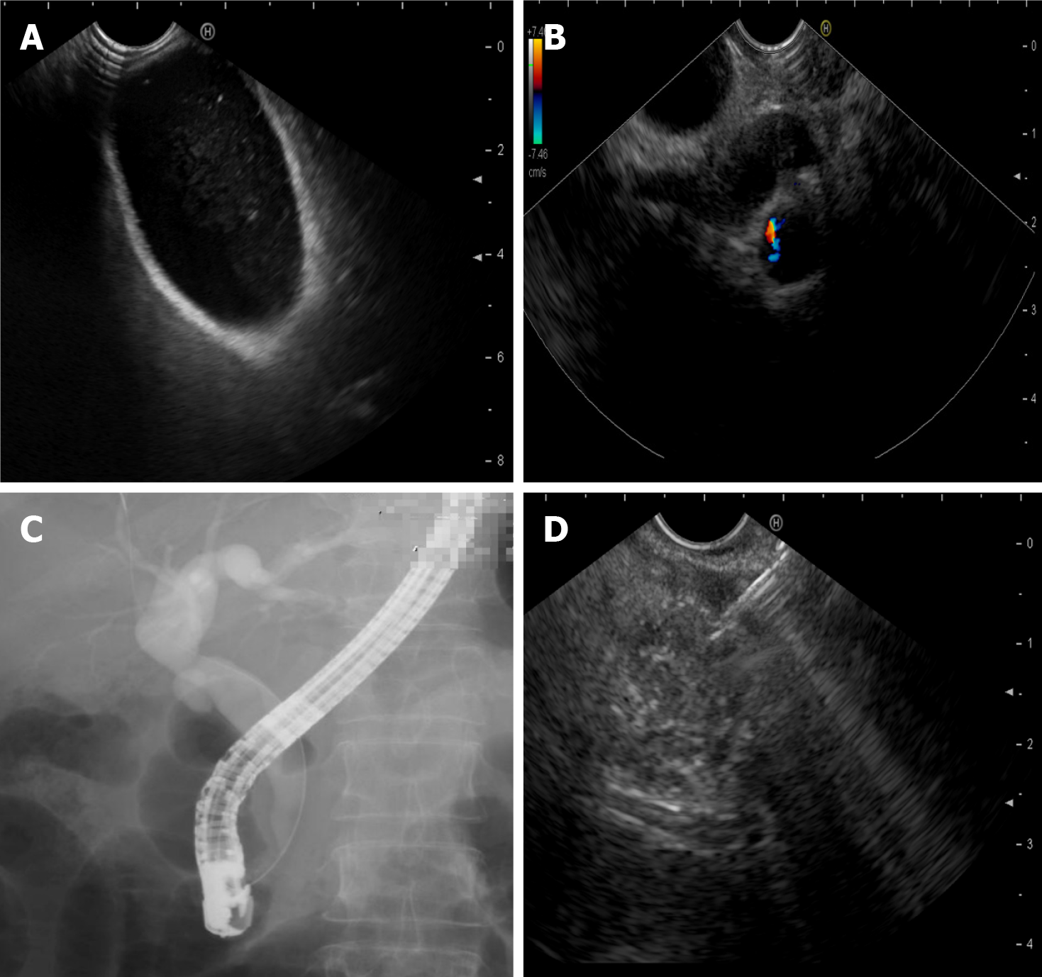
Figure 1 Endoscopically diagnosed etiology of pancreatitis.
A: Cholecystic microlithiasis detected by endoscopic ultrasound (EUS); B: Biliary sludge detected by EUS; C: Sphincter of Oddi dysfunction detected by endoscopic retrograde cholangiopancreatography; D: Small pancreatic cancer confirmed by EUS-guided fine-needle aspiration.
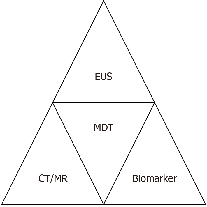
Figure 2 Structured pre-procedural evaluation for patients with infected necrotizing pancreatitis.
CT: Computed tomography; EUS: Endoscopic ultrasound; MDT: Multidisciplinary team; MR: Magnetic resonance.
Figure 3 Three-dimensional reconstruction model of infected necrotizing pancreatitis.
A: Three-dimensional reconstruction model of necrotic lesions; B: One transverse section of infected necrotizing pancreatitis; C: Transverse computed tomography section corresponding to Figure 2B.
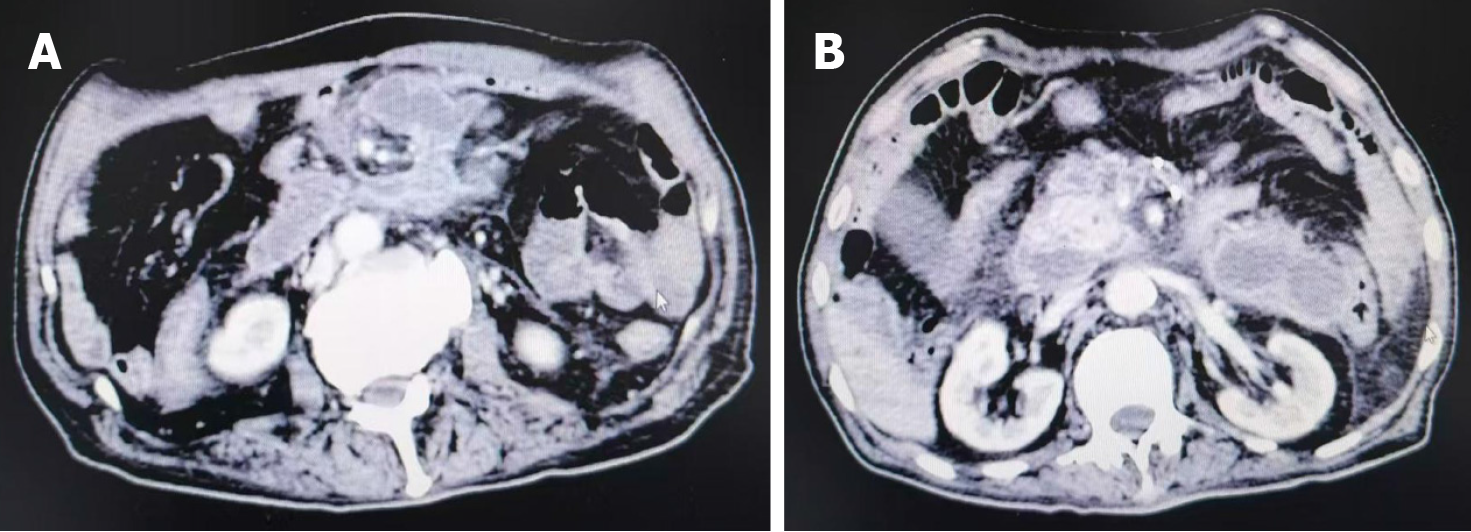
Figure 4 Post-percutaneous drainage fragmentations of necrotic collections.
A: Cross-sectional view of an infected necrotizing pancreatitis lesion after percutaneous drainage; B: Another view from the same post-procedural patient.
Figure 5 Graded balloon dilation.
A: Initial balloon dilation with a small-diameter balloon; B: Subsequent dilation with a large-diameter balloon.
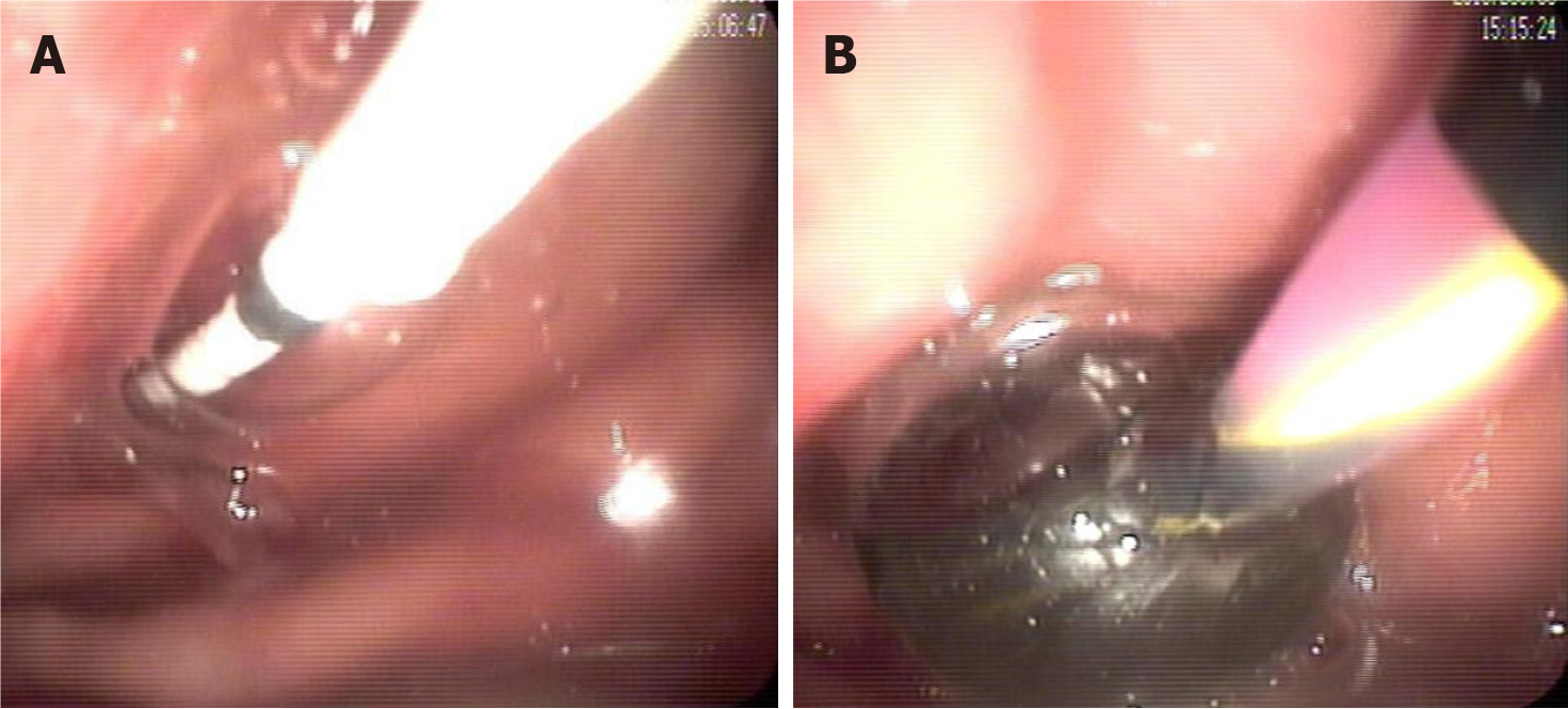
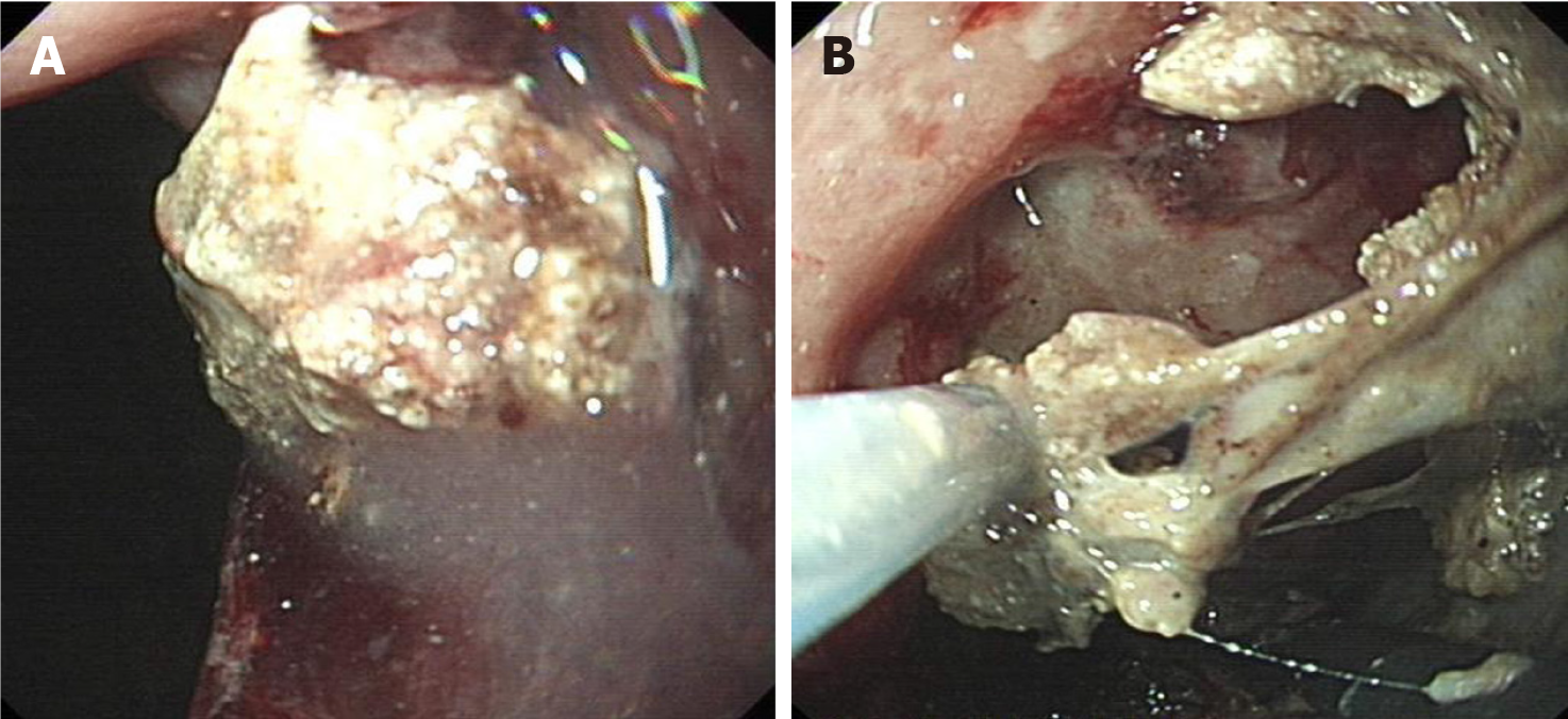
Figure 6 Endoscopic transgastric debridement of solid necrosis.
A: Transgastric drainage tract obstructed by solid necrosis in infected necrotizing pancreatitis; B: Endoscopic transgastric debridement.
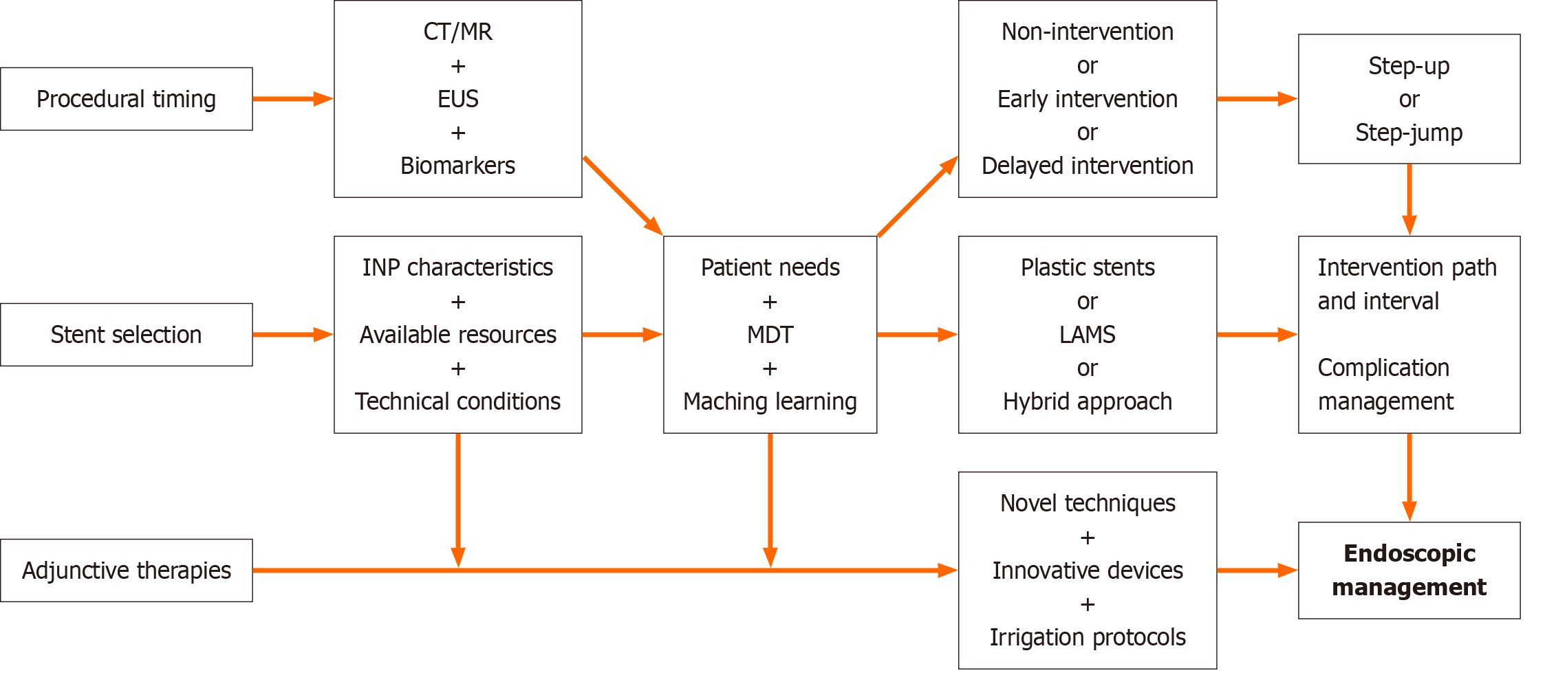
Figure 7 Endoscopic management flowchart of infected necrotizing pancreatitis.
CT: Computed tomography; EUS: Endoscopic ultrasound; INP: Infected necrotizing pancreatitis; LAMS: Lumen-apposing metal stents; MDT: Multidisciplinary team; MR: Magnetic resonance.
- Citation: Zeng Y, Zhang JW, Yang J. Endoscopic management of infected necrotizing pancreatitis: Advancing through standardization. World J Gastroenterol 2025; 31(20): 107451
- URL: https://www.wjgnet.com/1007-9327/full/v31/i20/107451.htm
- DOI: https://dx.doi.org/10.3748/wjg.v31.i20.107451


