Copyright
©The Author(s) 2025.
World J Gastroenterol. May 28, 2025; 31(20): 100192
Published online May 28, 2025. doi: 10.3748/wjg.v31.i20.100192
Published online May 28, 2025. doi: 10.3748/wjg.v31.i20.100192
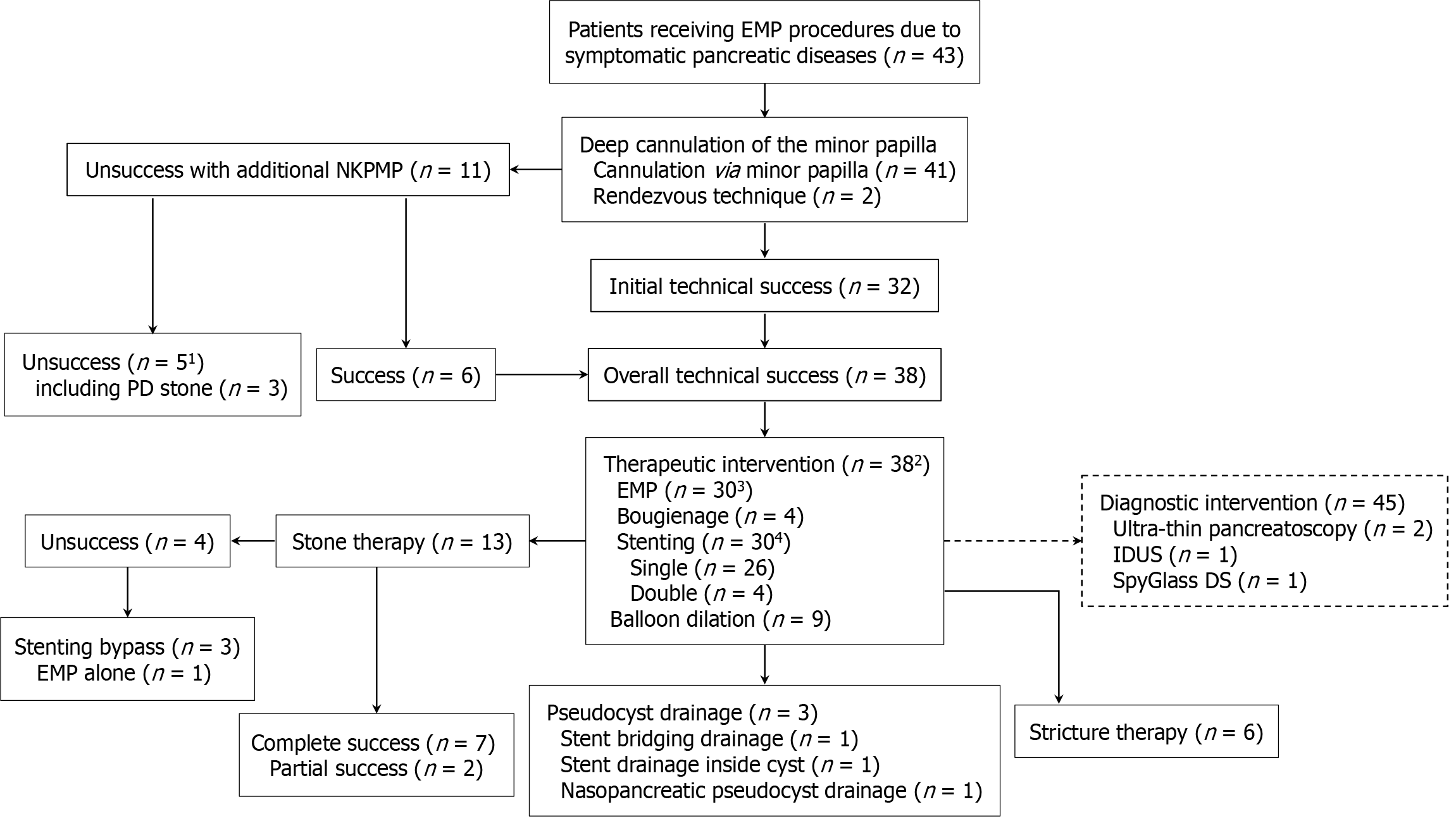
Figure 1 Technical details of endoscopic minor papilla intervention procedures in representative patients.
The solid line indicates therapeutic intervention and unsolid line indicates diagnostic intervention. 1Including 4 patients with obstructive chronic pancreatitis (3 of them with impacted stones) and 1 patient with pancreas divisum and chronic pancreatitis. 2Two or more interventional procedures were performed in some individual patients. 3Two patients received both needle-knife precut minor papillotomy (NKPMP) and endoscopic minor papillotomy (EMP). 4A 5 Fr stent (n = 10) or 7-8.5 Fr stent (n = 20) was used and ultimately exchanged in some cases with a single 10 Fr stent (n = 2) and double 7 Fr stents (n = 4) to treat the stricture. 5Four patients received diagnostic intervention after therapeutic intervention procedures including EMP (n = 4) and balloon dilation of the minor papilla and stricture (n = 1) for SpyGlass passing through the stricture and balloon sweep (n = 2) for mucin removal. IDUS: Intraductal ultrasound; PD: Pancreatic duct.
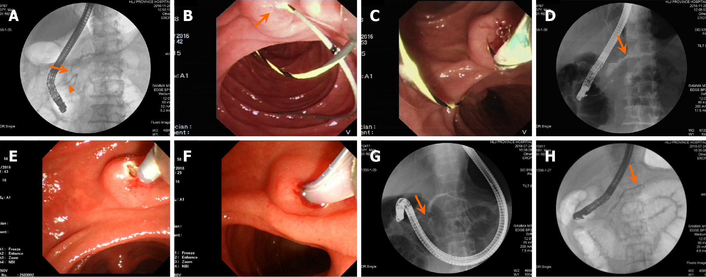
Figure 2 Illustration of the technical success of deep cannulation via the minor papilla in two patients.
A-D: Patient 1 receiving the rendezvous technique: Pancreatography shows the reverse-Z type of the main pancreatic duct (MPD) with a proximal stricture (arrow) of MPD, and the guidewire antegrade into the narrowed accessory pancreatic duct (arrowhead; A); Under duodenoscopy, the guidewire retrogradely enters from the major papilla and antegrade through the minor papilla (arrow) into the duodenum (B); The papillotome is inserted into the minor papilla over the other end of the guidewire pulled back from the biopsy channel (C); Pancreatic ductography demonstrates the guidewire (arrow) adjustment entering upstream of the MPD with successful deep cannulation (D); E-H: Patient 2 receiving additional free-hand needle-knife precut minor papillotomy: Endoscopic view shows the needle-knife precut minor papillotomy in progress (E); The papillotome is inserted into the minor papilla after incisions of the minor papilla (F); Pancreatic ductography displays a short type accessory pancreatic duct (APD) and a proximal stricture (arrow) of MPD, and upstream MPD dilation (G); and The guidewire (arrow) successfully passes through the APD into the upstream MPD, achieving successful deep cannulation (H).
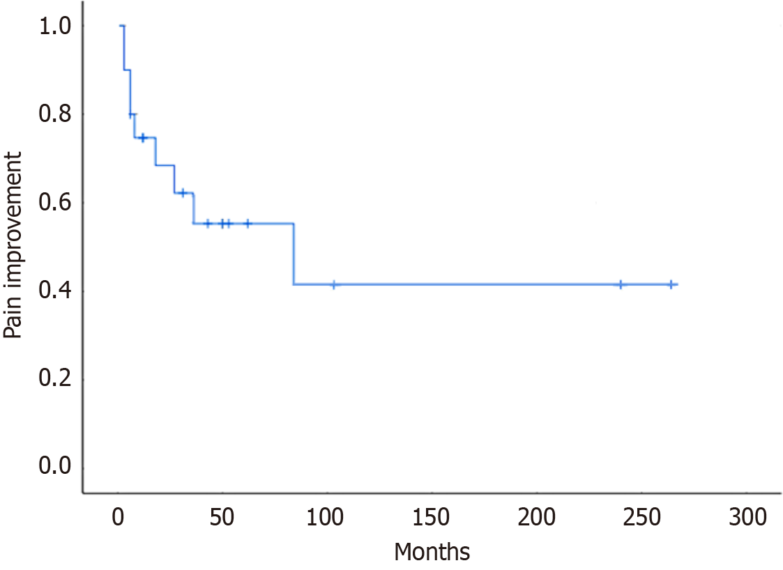
Figure 3 Kaplan-Meier curves showing the long-term clinical success (pain free) after endoscopic minor papilla intervention procedures.
Cumulative clinical success at 1 year, 3 years, and 7 years were 747%, 55.3%, and 41.5%, respectively, in 22 patients (19 with obstructive chronic pancreatitis and 3 with pancreas divisum) who underwent endotherapy) during a follow-up period of median 57.5 (7-266) months.
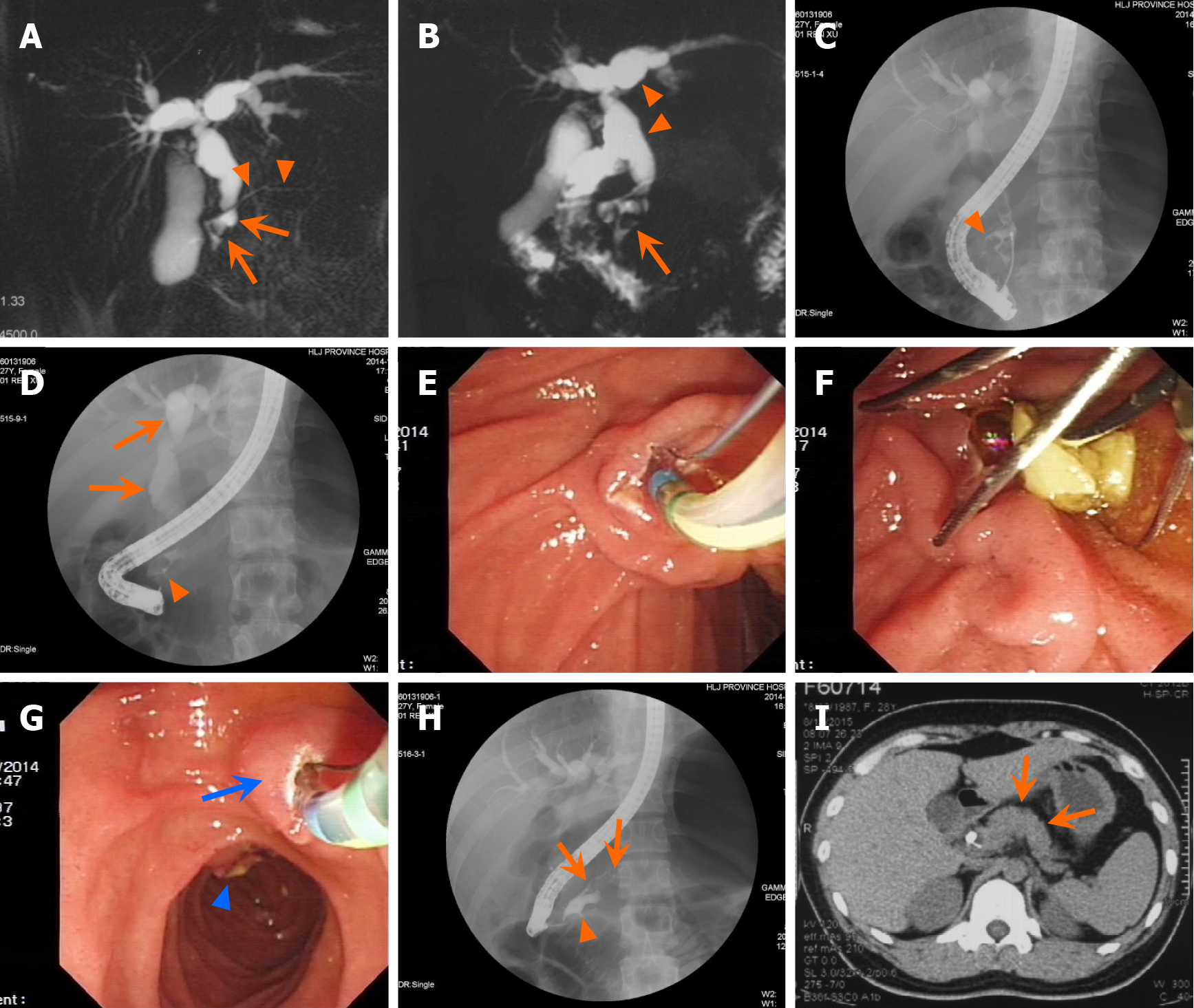
Figure 4 A case of complex pancreaticobiliary maljunction with complete pancreatic divisum and accompanied with stones in both the dorsal duct and the common bile duct.
A: Magnetic resonance cholangiopancreatography (MRCP) shows notable localized dilation of the dorsal duct (arrow) and a pancreatic stone (arrow), accompanied by a markedly narrowed upstream dorsal duct (arrowhead) and dilation of the intrahepatic and extrahepatic bile ducts. The ventral duct is not visualized; B: MRCP displays the presence of a common bile duct stone (arrow) and dilated dorsal duct, along with dilation of the intrahepatic and extrahepatic bile ducts (arrowhead); C: Cholangiography via the major papilla shows localized dilation of the dorsal duct and a pancreatic stone (arrowhead), indicating an anomalous junction between the dorsal duct and common bile duct; D: Cholangiography via the major papilla shows a large stone in the dorsal duct (8 mm × 12 mm; arrowhead), along with dilation of the intrahepatic and extrahepatic bile ducts progressing towards a choledochal cyst (arrow); E: Endoscopic sphincterotomy is being performed; F: The common bile duct stone is extracted using a basket; G: Endoscopic minor papillotomy (arrow) is being carried out and the major papilla can be seen in distance; H: Pancreatography via the minor papilla after removed the pancreatic stone reveals localized dilation of the dorsal duct (arrowhead) and a narrowed upstream dorsal duct (arrow); I: Computed tomography scan indicates no agenesis of the dorsal pancreas (i.e. absence of the pancreatic body and tail (arrow).
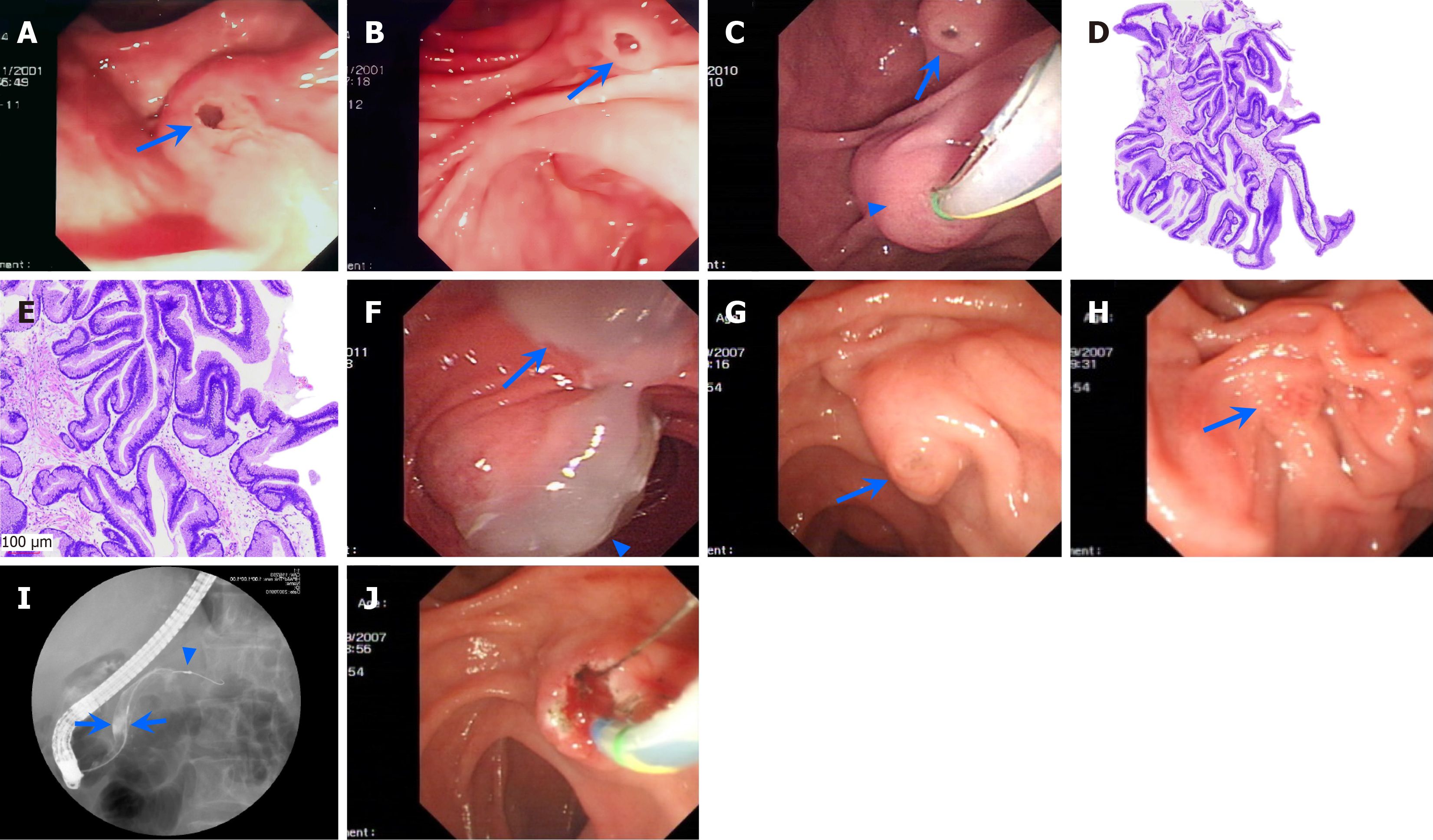
Figure 5 Illustrations of the branch duct-type and main duct-type intraductal papillary mucinous neoplasms in 3 patients.
A and B: Patient 1 with branch duct-type intraductal papillary mucinous neoplasm (IPMN): Duodenoscopy shows the patulous orifice of the major papilla with a fish eye-like appearance (arrow; A), and the patulous orifice of the minor papilla with a fish eye-like appearance (arrow) and mucus flowing out from the orifice before the patient underwent ultra-thin pancreatoscopy via the minor papilla (B); C-F: Patient 2 with main duct-type IPMN: Duodenoscopy shows the enlarged major papilla (arrowhead) with doing deep cannulation (but unsuccessful), and the patulous orifice of the minor papilla (arrow; C), histological examination shows villous papillary architecture composed of pseudostratified tall columnar cells with cigar-shaped enlarged nuclei and basophilic cytoplasm with a large amount of apical mucin, and low-grade and intestinal-type IPMN (hematoxylin and eosin staining, 40 × and 100 ×, respectively; D and E), and a large amount of gelatinous mucous flowing out from both the major (arrowhead) and minor papilla (arrow) 10 months after an endoscopic minor papilla intervention procedure (F); G-J: Patient 3 with main duct-type IPMN: Duodenoscopy shows the enlarged minor papilla (arrow), with mucus visible at the opening (G), the major papilla (arrow) of normal size, with a narrower opening (failed deep cannulation (H), and pancreatic ductography reveals notably dilated (13 mm) dorsal duct (arrows) without filling defects (arrowhead) in the dorsal ducts of the pancreatic body and tail, and the ventral duct (I), and endoscopic minor papillotomy is being performed by using a pull-type papillotome (J).
- Citation: Ren X, Qu YP, Xia T, Tang XF. Technical success, clinical efficacy, and safety of endoscopic minor papilla interventions for symptomatic pancreatic diseases. World J Gastroenterol 2025; 31(20): 100192
- URL: https://www.wjgnet.com/1007-9327/full/v31/i20/100192.htm
- DOI: https://dx.doi.org/10.3748/wjg.v31.i20.100192


