©The Author(s) 2025.
World J Gastroenterol. Mar 28, 2025; 31(12): 102423
Published online Mar 28, 2025. doi: 10.3748/wjg.v31.i12.102423
Published online Mar 28, 2025. doi: 10.3748/wjg.v31.i12.102423
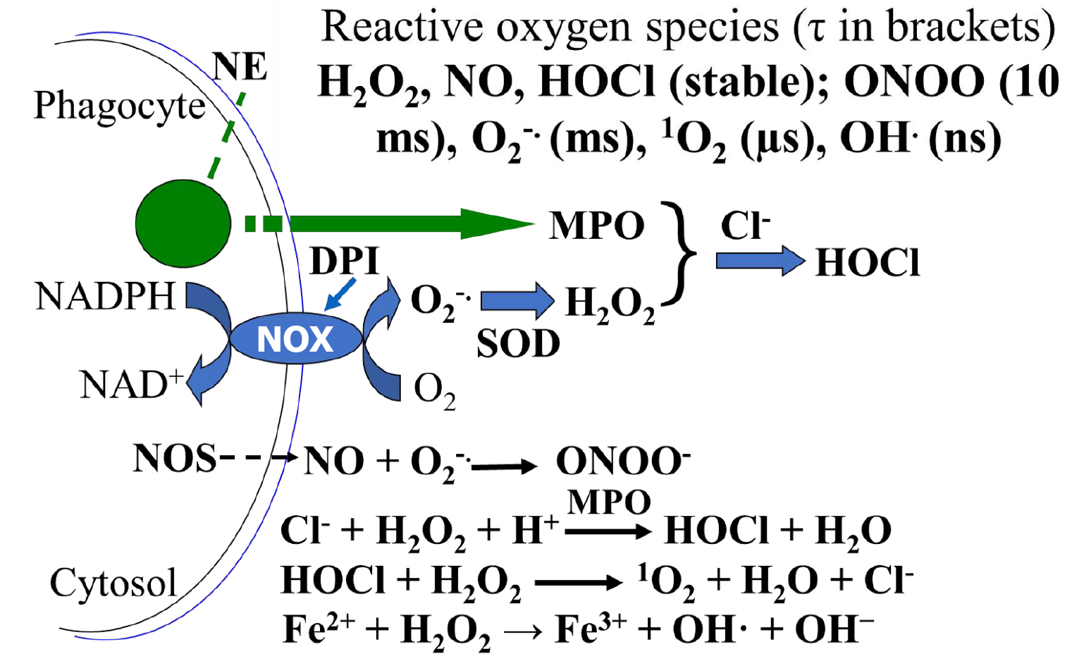
Figure 1 Reactive oxygen species and their average lifetime.
The nicotinamide adenine dinucleotide phosphate oxidase of phagocytes catalyzes the generation and release of superoxide (O2-.) after transfer of an electron to molecular oxygen (O2), O2-. is transformed into hydrogen peroxide (H2O2) by superoxide dismutase. H2O2 reacts with chloride anion, a major anion in the extracellular fluid, catalyzed by myeloperoxidase, to produce hypochlorous acid (HOCl), a bactericidal chemical. Nitric oxide (NO) produced by NO synthase reacts with O2-. to generate peroxynitrite anion. HOCl reacts with H2O2 to generate delta singlet oxygen (1O2). Ferrous iron reacts with H2O2 in Fenton reaction to produce ferric ion and hydroxyl radical (OH.). Note that H2O2, NO, HOCl are relatively stable, but peroxynitrite anion, O2-., 1O2, OH., has decreasing lifetimes of 10 ms, ms, μs, and ns respectively. 1O2 shows specificity in oxidative reactions (than OH-, for example) and has a diffusion distance from 10 nm to 150 nm, depending on the site of generation and methods of measurement and detection. NE: Neutrophil elastase; NADPH: Nicotinamide adenine dinucleotide phosphate; NAD: Nicotinamide adenine dinucleotide; NOS: Nitric oxide synthase; NO: Nitric oxide; H2O2: Hydrogen peroxide; HOCl: Hypochlorous acid; ONOO: Peroxynitrite anion; O2-.: Superoxide; 1O2: Delta singlet oxygen; OH.: Hydroxyl radical; DPI: Diphenyleneiodonium chloride; MPO: Myeloperoxidase; NOX: Nicotinamide adenine dinucleotide phosphate oxidase; Cl-: Chloride anion; SOD: Superoxide dismutase; H+: Proton; Fe2+: Ferrous iron; Fe3+: Ferric ion.
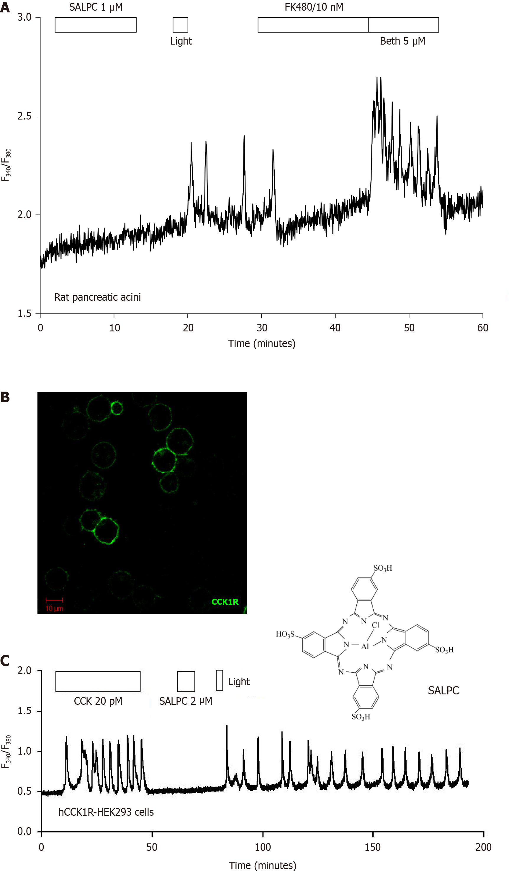
Figure 2 Permanent photodynamic activation of cholecystokinin 1 receptor.
A: Photodynamic action with sulphonated aluminum phthalocyanine (SALPC) (1 μmol/L; light λ > 580 nm, 53000 Lux/ 72.4 mW/cm2) triggered persistent calcium oscillations in perifused rat pancreatic acini loaded with Fura-2 AM, which were completely blocked by cholecystokinin 1 receptor (CCK1R) antagonist FK480 (10 nM); after blockade by FK480, the muscarinic agonist bethanechol (5 μmol/L) could still elicit a robust calcium response. Modified from An et al[28]. Note that the photosensitizer SALPC was exposed to perifused rat pancreatic acini only briefly (10 minutes) then washed out, only residue photosensitizer remaining on the acinar cell plasma membrane was then irradiated by red light. Reproduced from An et al[28]; B: The human CCK1R (green) was expressed in HEK293 cells, as detected by immunocytochemsitry. Scale bar: 10 μm. Modified from Jiang et al[13]; C: The positively expressing human CCK1R-HEK293 cells were loaded with fluorescent calcium indicator Fura-2 AM, perifused, CCK 20 pM, SALPC 2 μmol/L and light (λ > 580 nm, 20500 Lux/ 36.7 mW/cm2) were applied as indicated. Note the reversible calcium oscillations with CCK 20 pM stimulation, but persistent calcium oscillations after photodynamic action with SALPC. Modified from Jiang et al[13]. Note that the photosensitizer SALPC was exposed to perifused human CCK1R-HEK293 cells only for 10 minutes before wash out. Only residual photosensitizer SALPC on the plasma membrane was irradiated by red light (> 580 nm). Note that the peripheral sulphate groups (4) of SALPC structure ensure aqueous solubility and little ability to enter the cell interiors, enabling plasma membrane specificity. Reproduced from Jiang et al[13]. SALPC: Sulphonated aluminum phthalocyanine; hCCK1R: Human cholecystokinin 1 receptor. Citation for Figure 2A: An YP, Xiao R, Cui H, Cui ZJ. Selective activation by photodynamic action of cholecystokinin receptor in the freshly isolated rat pancreatic acini. Br J Pharmacol 2003; 139: 872-880. Copyright© Nature Publishing Group 2003. Published by John Wiley and Sons; Citation for Figure 2B and C: Jiang HN, Li Y, Jiang WY, Cui ZJ. Cholecystokinin 1 Receptor - A Unique G Protein-Coupled Receptor Activated by Singlet Oxygen (GPCR-ABSO). Front Physiol 2018; 9: 497. Copyright© The Authors 2018. Published by Frontiers Media S.A. The authors have obtained the permission for figure using (Supplementary material).
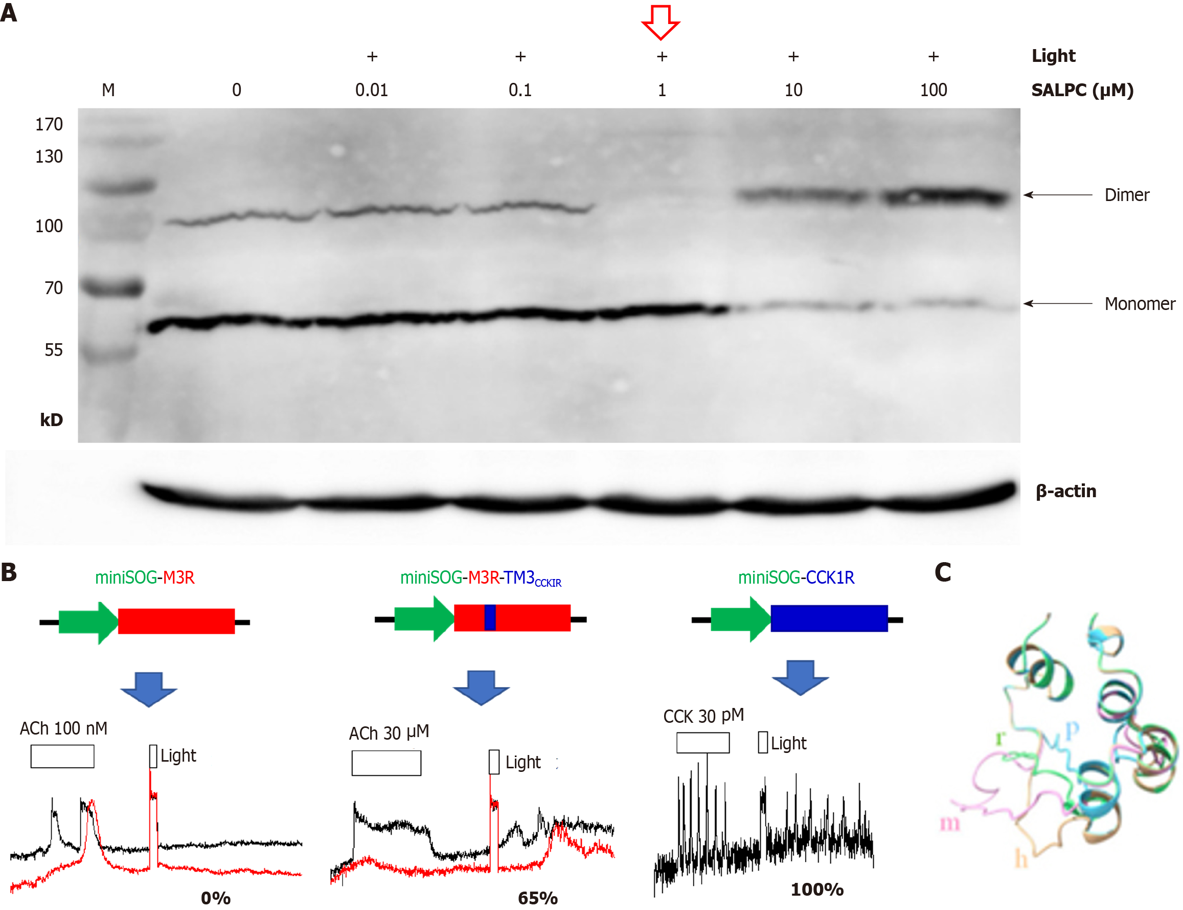
Figure 3 Molecular mechanisms for permanent photodynamic activation of cholecystokinin 1 receptor.
A: Near quantitative photodynamic conversion [at sulphonated aluminum phthalocyanine (SALPC) 1 μmol/L + light, an intensity that would activate cholecystokinin 1 receptor (CCK1R) in live cells, note the red arrow] of CCK1R from dimer to monomer. Membrane proteins isolated from rat pancreatic acini were subject to photodynamic action (SALPC 0.01 to 100 μmol/L, light λ > 580 nm, 36.7 mW/cm2) before Western blot analysis. Note that higher photodynamic doses (SALPC 10 μmol/L, 100 μmol/L + light), not used in live cell experiments, led to cross-linking of CCK1R monomer. Modified from Jiang et al[41]; B: Transplantation of transmembrane domain 3 (TM3) from CCK1R to type 3 muscarinic acetylcholine receptor (M3R) generates chimeric receptor mini singlet oxygen generator (miniSOG)-M3R-TM3CCK1R, rendering non-susceptible M3R photodynamically activated. Calcium tracings are marked underneath with averaged percentage receptor activation (miniSOG-M3R 0%, miniSOG-tagged chimeric receptor 65%) compared with miniSOG-CCK1R (100%). Note that miniSOG (green arrow) is tagged to the N-terminus of M3R (red bar), CCK1R (blue bar), or the chimera M3R-TM3CCK1R (TM3CCK1R portion shown as blue and M3R portion shown as red). For clarity, only 2 cells are shown in the left and middle panel, and 1 cell is shown in the right panel. Reproduced from Li and Cui[43]; C: The length of secondary structure-free region in intracellular loop 3 (ICL3) correlates with sensitivity to photodynamic CCK1R activation: Mouse > rat > Peking duck. The complete ICL3 is shown. Note the complete alignment at beginning (top left) and end of ICL3. Alphabets are color coded on structure (m: mouse; h: human; r: rat; p: Peking duck). Reproduced from Wang and Cui[32]. SALPC: Sulphonated aluminum phthalocyanine; CCK1R: Cholecystokinin 1 receptor; M3R: Type 3 muscarinic acetylcholine receptor; miniSOG: Mini singlet oxygen generator; TM3: Transmembrane domain 3. Citation for Figure 3A: Jiang WY, Li Y, Li ZY, Cui ZJ. Permanent Photodynamic Cholecystokinin 1 Receptor Activation: Dimer-to-Monomer Conversion. Cell Mol Neurobiol 2018; 38: 1283-1292. Copyright© Springer Science Business Media 2018. Published by Springer Nature; Citation for Figure 3B: Li Y, Cui ZJ. Transmembrane Domain 3 Is a Transplantable Pharmacophore in the Photodynamic Activation of Cholecystokinin 1 Receptor. ACS Pharmacol Transl Sci 2022; 5: 539-547. Copyright© American Chemical Society 2022. Published by American Chemical Society; Citation for Figure 3C: Wang J, Cui ZJ. Photodynamic Activation of Cholecystokinin 1 Receptor Is Conserved in Mammalian and Avian Pancreatic Acini. Biomedicines 2023; 11: 885. Copyright© The Authors 2023. Published by MDPI. The authors have obtained the permission for figure using (Supplementary material).
- Citation: Cui ZJ. To activate a G protein-coupled receptor permanently with cell surface photodynamic action in the gastrointestinal tract. World J Gastroenterol 2025; 31(12): 102423
- URL: https://www.wjgnet.com/1007-9327/full/v31/i12/102423.htm
- DOI: https://dx.doi.org/10.3748/wjg.v31.i12.102423


