Copyright
©The Author(s) 2022.
World J Gastroenterol. Apr 21, 2022; 28(15): 1536-1547
Published online Apr 21, 2022. doi: 10.3748/wjg.v28.i15.1536
Published online Apr 21, 2022. doi: 10.3748/wjg.v28.i15.1536
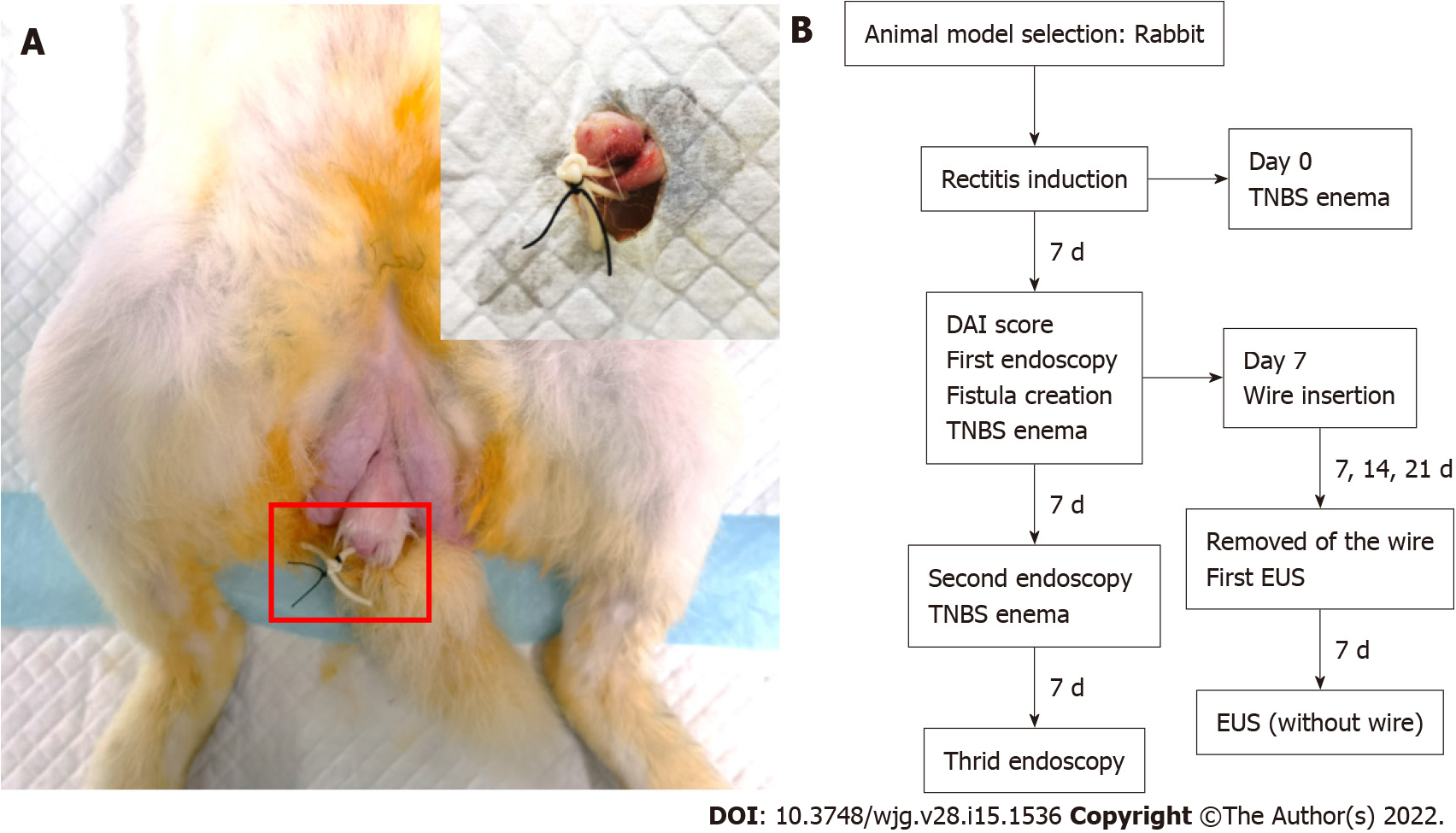
Figure 1 Photograph of the external opening of the anal fistula, which was approximately 1 cm away from the anus, and a flowchart of the study protocol.
A: The rubber band was used to hang the seton, and the leather band was fixed with a No. 0 operation seton to prevent slippage; B: One rabbit died one week after surgery. The remaining 17 rabbits were marked according to the length of insertion time. Three rabbits each were randomly selected from groups A and B. TNBS: 2,4,6-Trinitrobenzene sulfonic acid; DAI: Disease activity index; EUS: Endoscopic ultrasonography.
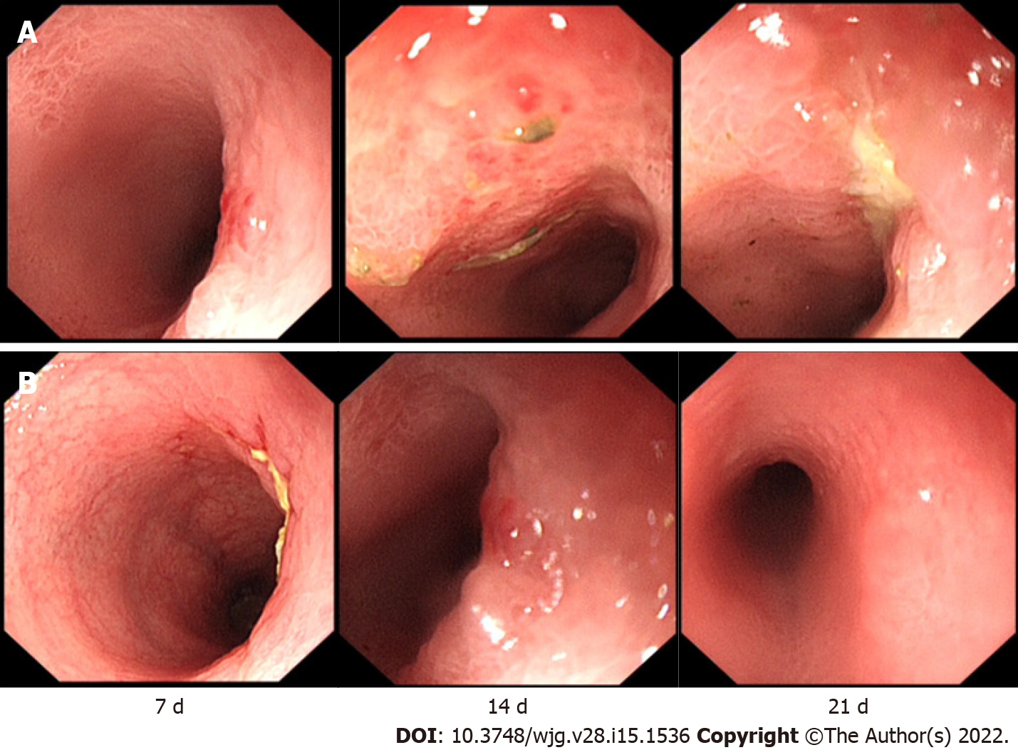
Figure 2 The rabbits in groups A and B underwent endoscopy 3 times.
A: In group A, mild mucosal erosion was observed on the 7th day. On the 14th day, a large area of mucosal oedema and erosion appeared. On the 21st day, the surrounding mucosa was swollen, and a central ulcer had formed; B: In group B, ulceration occurred on the 7th day. On the 14th day, the mucosa was swollen, and the surface was hyperaemic and eroded. On the 21st day, the main manifestation was local congestion.
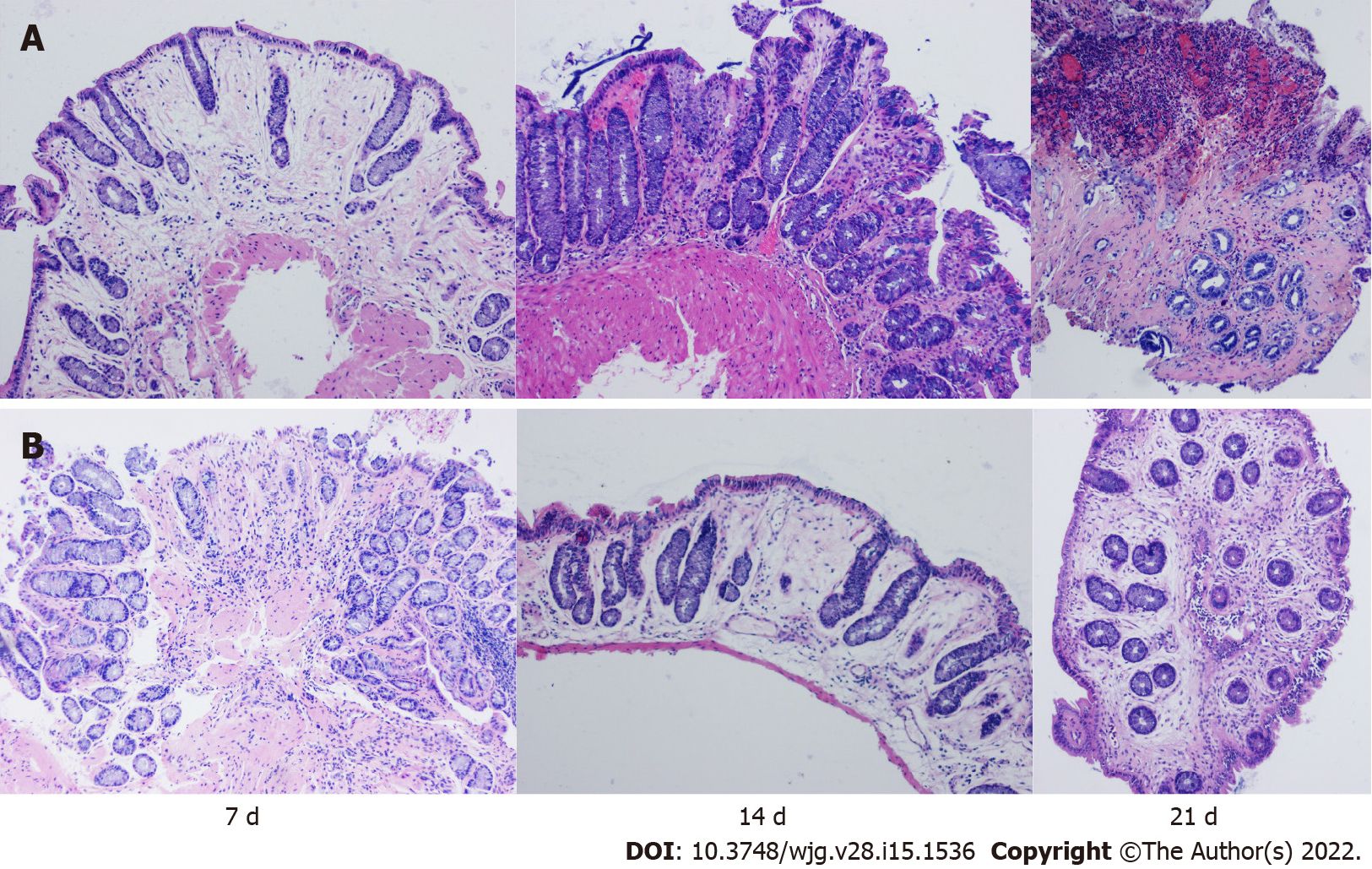
Figure 3 The rabbits in groups A and B underwent three consecutive endoscopic biopsies, and samples were stained with haematoxylin-eosin.
A: Group A exhibited mild to moderate inflammation on day 7, manifested by fewer crypts, marked mucosal oedema, and minimal inflammatory cell infiltration; Group A exhibited fibrosis, hyperaemia, and moderate inflammatory cell infiltration on day 14, and severe inflammatory mucosal ulceration on day 21; B: On the 7th day, there was a higher number of shallow crypts, less oedema and less inflammatory cell infiltration in group B. On the 14th day, there was severe oedema, decreased crypts and less inflammatory cell infiltration in the mucosa, and on the 21st day, there was moderate fibrosis and oedema, mild inflammatory cell infiltration and a complete crypt structure (magnification ×100).
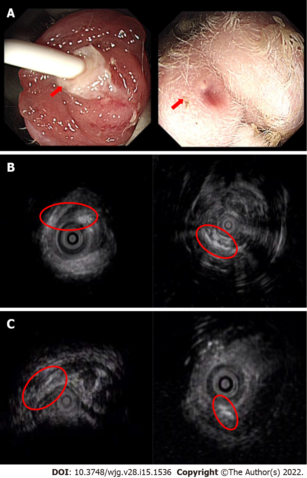
Figure 4 Visualization of a transsphincteric anal fistula via endoscopic ultrasonography.
A: The internal and external openings of the experimental fistula can be directly observed; B: All rabbits with a 21-d insertion time of the surgical thread showed a complete fistula; C: The rabbits with short thread insertion times had different degrees of fistula healing or the disappearance of internal and external fistulas. D0: Day 0; D7: Day 7.
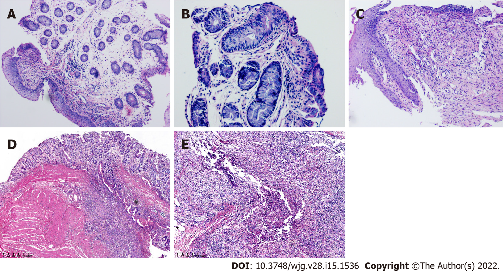
Figure 5 The histological characteristics of a fistula tract.
A: Magnification ×100; B: Magnification ×200; C: Magnification ×100. The early histological changes of fistula are shown; D and E: Longitudinal sections; histological results (rabbits with setons inserted 21 d) showing the inflamed fistula tract. The fistula lumen is visible with internal (digestive side) and external (perineal skin with adipocytes) orifices. There were local inflammatory signs of suppurative inflammation with abscess formation around the fistula.
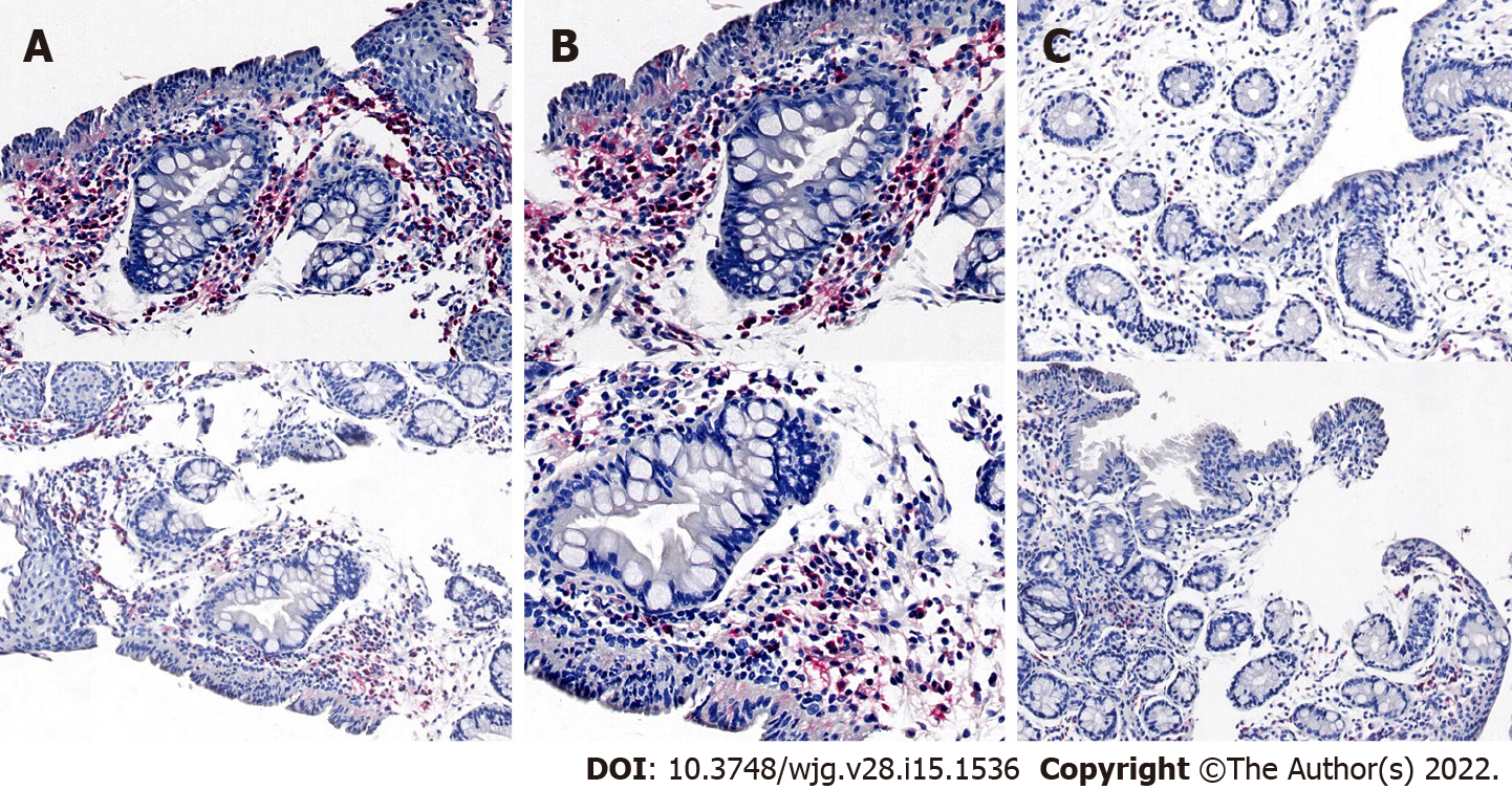
Figure 6 Immunohistochemistry of a fistula tract.
A: Magnification ×10; B: Magnification ×20; C: Control. Immunohistochemistry of the fistula tract using anti-CD68 antibodies (upper panel) or anti-MPO antibodies (lower panel).
- Citation: Lu SS, Liu WJ, Niu QY, Huo CY, Cheng YQ, Wang EJ, Li RN, Feng FF, Cheng YM, Liu R, Huang J. Establishing a rabbit model of perianal fistulizing Crohn’s disease. World J Gastroenterol 2022; 28(15): 1536-1547
- URL: https://www.wjgnet.com/1007-9327/full/v28/i15/1536.htm
- DOI: https://dx.doi.org/10.3748/wjg.v28.i15.1536


