©The Author(s) 2021.
World J Gastroenterol. Oct 28, 2021; 27(40): 6825-6843
Published online Oct 28, 2021. doi: 10.3748/wjg.v27.i40.6825
Published online Oct 28, 2021. doi: 10.3748/wjg.v27.i40.6825
Figure 1 A deep convolutional network.
An example deep learning segmentation network, the U-Net. A series of convolutions, combined with downsampling and upsampling to learn feature maps at different scales, are used to output a segmentation map. An example of a convolution, and feature map are shown on the right.
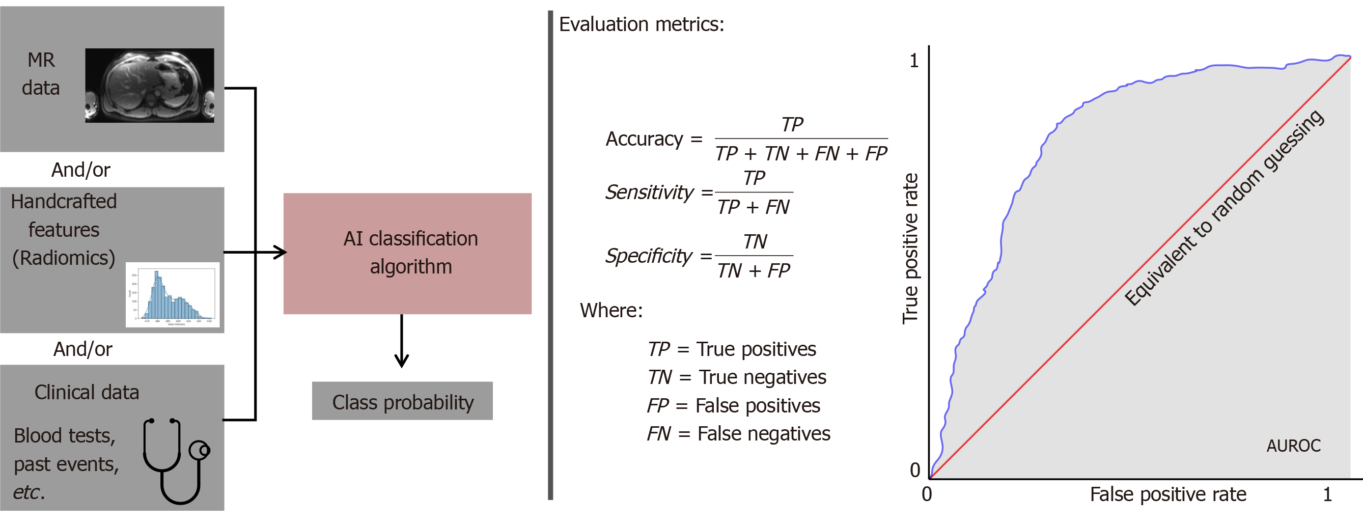
Figure 2 Classification algorithms and their performance metrics.
Artificial intelligence classification algorithms use the combination of data provided to them and output a class probability. They are often evaluated according to the metrics on the right. MR: Magnetic resonance; AUROC: Area under the receiver operating characteristic curve.
- Citation: Hill CE, Biasiolli L, Robson MD, Grau V, Pavlides M. Emerging artificial intelligence applications in liver magnetic resonance imaging. World J Gastroenterol 2021; 27(40): 6825-6843
- URL: https://www.wjgnet.com/1007-9327/full/v27/i40/6825.htm
- DOI: https://dx.doi.org/10.3748/wjg.v27.i40.6825


