Copyright
©The Author(s) 2021.
World J Gastroenterol. May 21, 2021; 27(19): 2299-2311
Published online May 21, 2021. doi: 10.3748/wjg.v27.i19.2299
Published online May 21, 2021. doi: 10.3748/wjg.v27.i19.2299
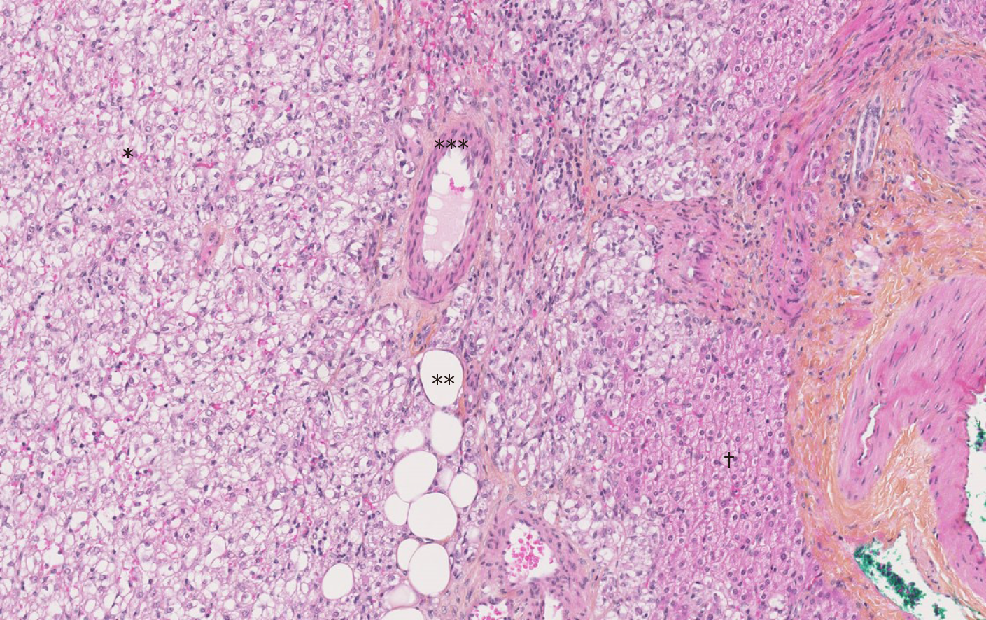
Figure 1 The Hematoxylin-Eosin-Saffron staining image of hepatic angiomyolipoma.
There are three components of hepatic angiomyolipoma: vessel (*), adipocytes (**) and numerous epithelioid cells (***). There are fewer hepatocytes (†) (magnification × 10).
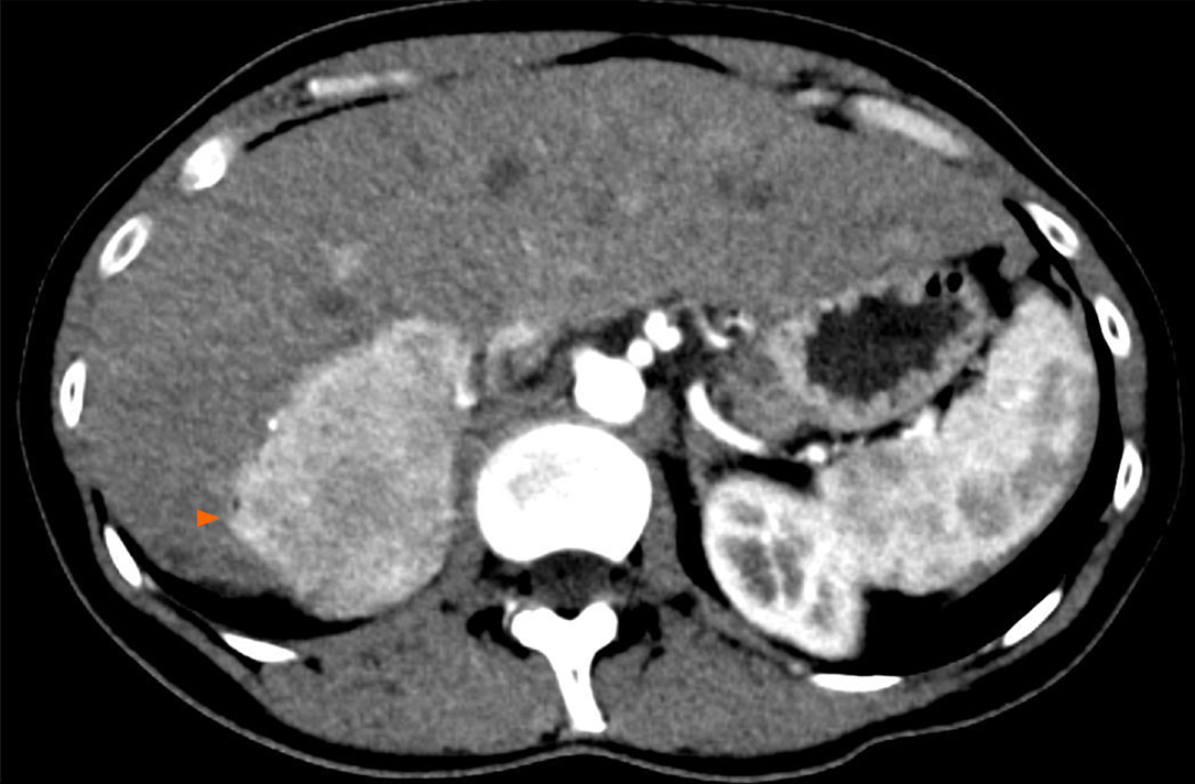
Figure 2 Angiomyolipoma in a healthy 33-year-old woman.
Abdominal computed tomography on arterial phase showed a hypervascular solid tumor localized in the right posterior segment (arrowheads).
Figure 3 T1 weighted magnetic resonance images.
Signal dropout at the periphery of the lesion due to fat contingents (arrowhead). A: In-phase; B: Opposed-phase.
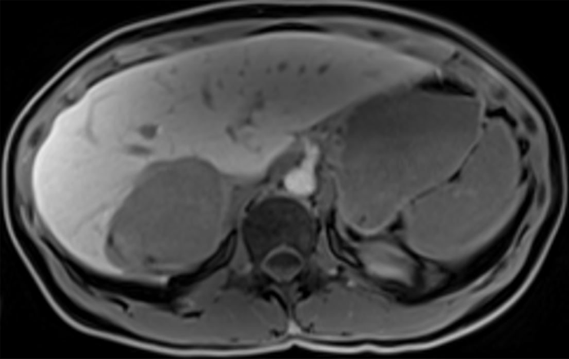
Figure 4 T1 weighted images one hour after hepatocyte-specific agent injection (gadobenate dimeglumine).
Hyposignal of the lesion indicates that this is not a hepatocytic tumor.
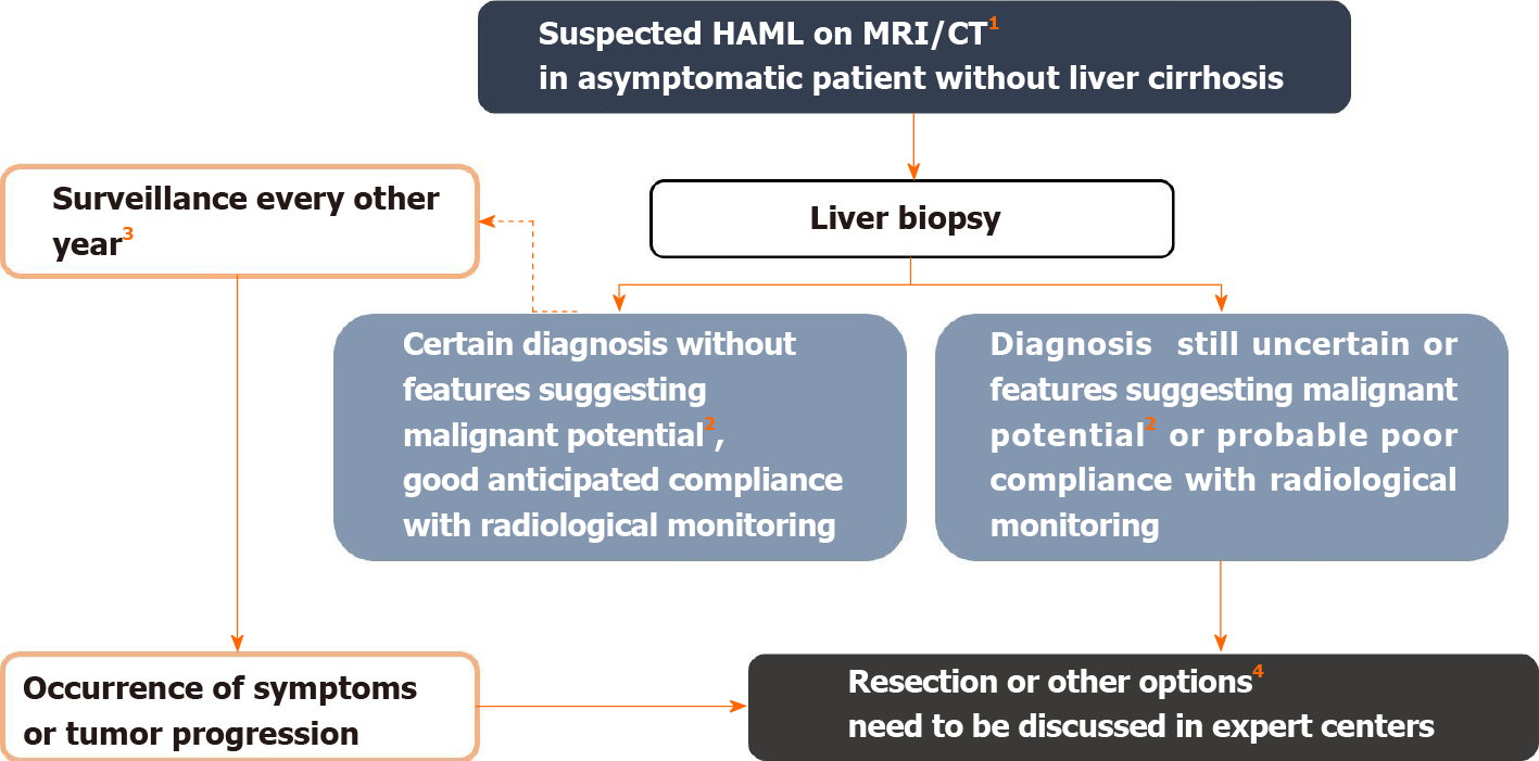
Figure 5 Management algorithm for suspected hepatic angiomyolipoma on imaging.
1Hepatic angiomyolipoma diagnosis is suggested in the presence of fatty tissue within the solid lesion or presence of wash-out. In the presence of tumor-related symptoms, surgical resection is considered first. 2Features suggesting malignant potential are reported in Table 2. Some authors also recommend surgery in the case of epithelioid-type hepatic angiomyolipoma, which would be at greater risk of progression. Likewise, an association with tuberous sclerosis complex is a condition that increases the risk of malignant transformation, by analogy with renal angiomyolipoma[3]. 3Monitoring maintained despite the benign nature of the initial diagnosis because the aggressive behavior of the tumor is difficult to predict. 4Other possible therapeutic options include mTOR inhibitors, radiofrequency ablation, arterial embolization in cases of hemorrhagic rupture, and liver transplantation. Citation: Klompenhouwer AJ, Verver D, Janki S, Bramer WM, Doukas M, Dwarkasing RS, de Man RA, IJzermans JNM. Management of hepatic angiomyolipoma: A systematic review. Liver Int 2017; 37(9): 1272-1280. Copyright ©The Author(s) 2017. Published by John Wiley and Sons[3]. MRI: Magnetic resonance imaging; CT: Computed tomography.
- Citation: Calame P, Tyrode G, Weil Verhoeven D, Félix S, Klompenhouwer AJ, Di Martino V, Delabrousse E, Thévenot T. Clinical characteristics and outcomes of patients with hepatic angiomyolipoma: A literature review. World J Gastroenterol 2021; 27(19): 2299-2311
- URL: https://www.wjgnet.com/1007-9327/full/v27/i19/2299.htm
- DOI: https://dx.doi.org/10.3748/wjg.v27.i19.2299


