©The Author(s) 2020.
World J Gastroenterol. Feb 21, 2020; 26(7): 770-776
Published online Feb 21, 2020. doi: 10.3748/wjg.v26.i7.770
Published online Feb 21, 2020. doi: 10.3748/wjg.v26.i7.770
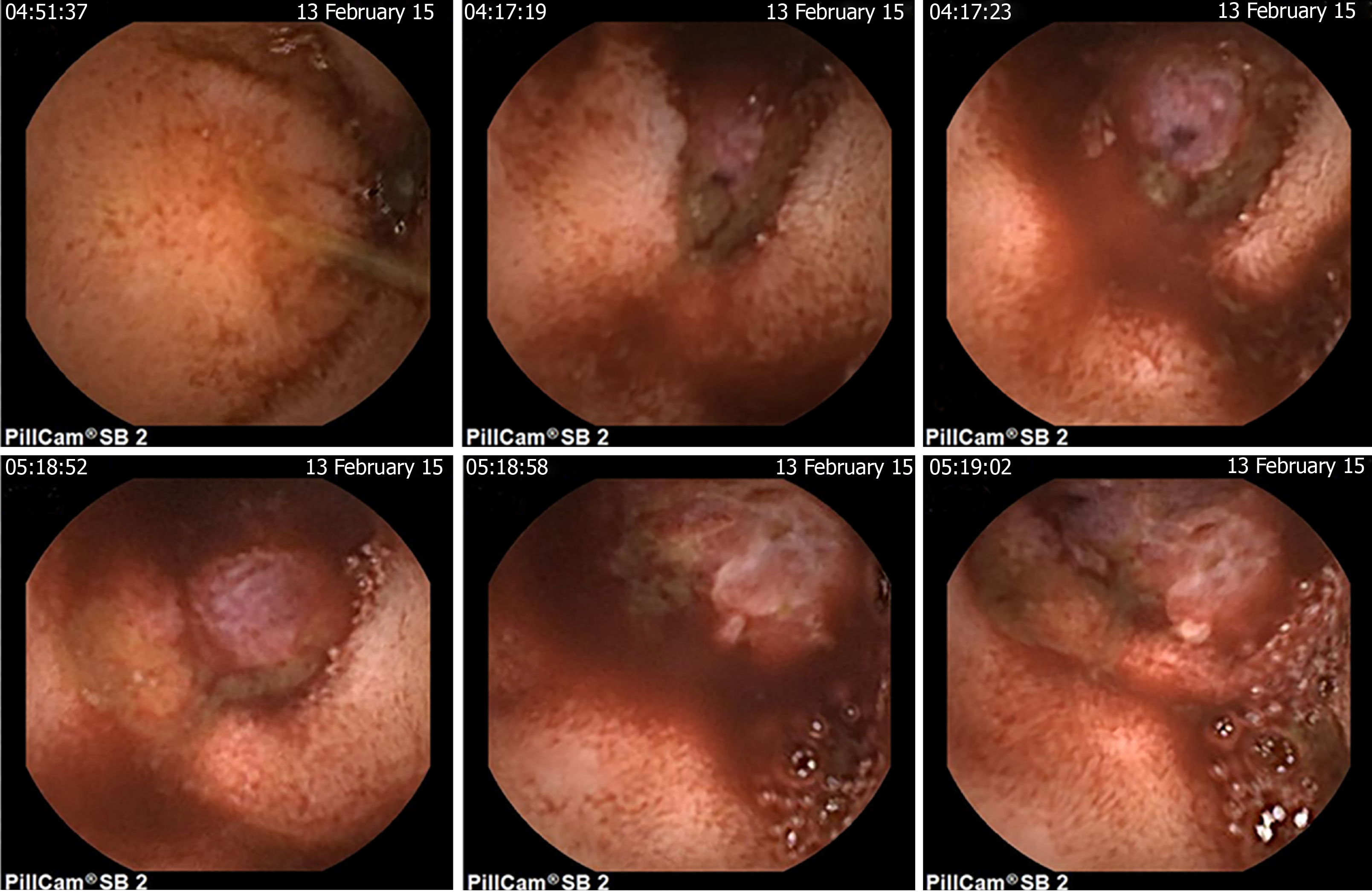
Figure 1 Capsule endoscopic characteristics of the intestinal glomus tumor from different perspectives.
Figure 2 Histological characteristics of the malignant glomus tumor in ileum.
A: Spindled tumor cells with branched or dilated vessels surrounded [hematoxylin and eosin (H&E) stain, 100 ×]; B: Spindled cells with high mitotic activity and nuclear atypia marked with arrows (H&E stain, 200 ×); and C: Tumor cells with vascular invasion (H&E stain, 100 ×).
Figure 3 Immunohistochemical staining characteristics of the malignant glomus tumor in ileum.
A: Smooth muscle actin; B: Vimentin; C: Caldesmon; and D: Ki-67.
- Citation: Chen JH, Lin L, Liu KL, Su H, Wang LL, Ding PP, Zhou Q, Liu H, Wu J. Malignant glomus tumor of the intestinal ileum with multiorgan metastases: A case report and review of literature. World J Gastroenterol 2020; 26(7): 770-776
- URL: https://www.wjgnet.com/1007-9327/full/v26/i7/770.htm
- DOI: https://dx.doi.org/10.3748/wjg.v26.i7.770


