©The Author(s) 2020.
World J Gastroenterol. Oct 7, 2020; 26(37): 5693-5704
Published online Oct 7, 2020. doi: 10.3748/wjg.v26.i37.5693
Published online Oct 7, 2020. doi: 10.3748/wjg.v26.i37.5693
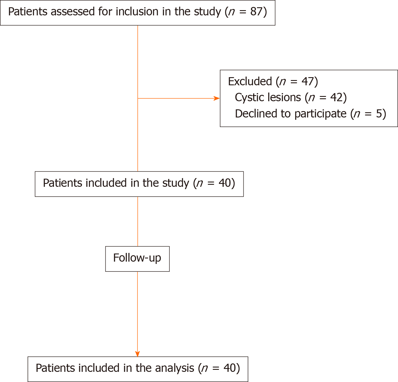
Figure 1 Study flow diagram.
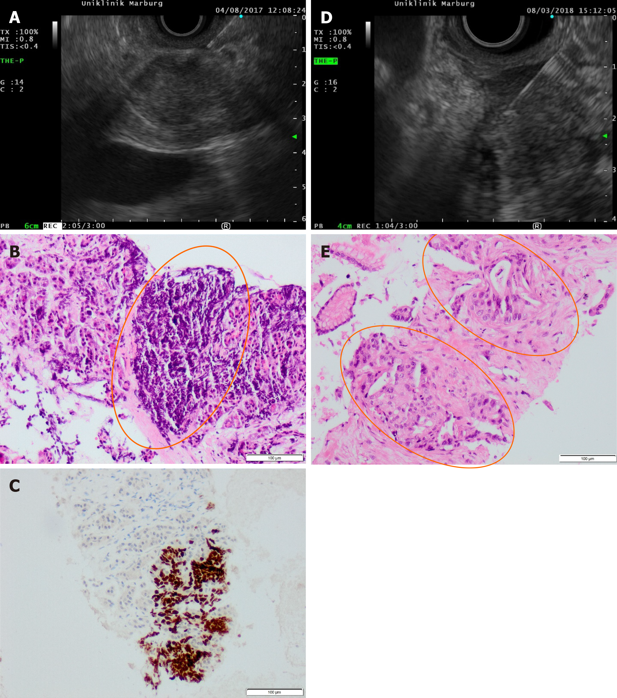
Figure 2 Endoscopic ultrasound and histology images of two lesions.
A: Endoscopic ultrasound-fine needle biopsies (EUS-FNB) of a pancreatic metastasis of a non-small-cell lung cancer; B: The corresponding hematoxylin-eosin (H&E) staining (100 ×), showing a cluster of small tumor cells, high nuclear to cytoplasm ratio and crush artifacts; C: The immunohistochemistry for TTF-1 reactive in the nuclei of tumor. D: EUS-FNB of a pancreatic adenocarcinoma with the Franseen needle; E: The corresponding H&E staining (100 ×), showing characteristic infiltrating glands with tumor cells with nuclear hyperchromatism, pleiomorphism and prominent nucleoli in a cell block.
- Citation: Stathopoulos P, Pehl A, Breitling LP, Bauer C, Grote T, Gress TM, Denkert C, Denzer UW. Endoscopic ultrasound-fine needle biopsies of pancreatic lesions: Prospective study of histology quality using Franseen needle. World J Gastroenterol 2020; 26(37): 5693-5704
- URL: https://www.wjgnet.com/1007-9327/full/v26/i37/5693.htm
- DOI: https://dx.doi.org/10.3748/wjg.v26.i37.5693


