Copyright
©The Author(s) 2020.
World J Gastroenterol. Jun 14, 2020; 26(22): 3000-3011
Published online Jun 14, 2020. doi: 10.3748/wjg.v26.i22.3000
Published online Jun 14, 2020. doi: 10.3748/wjg.v26.i22.3000
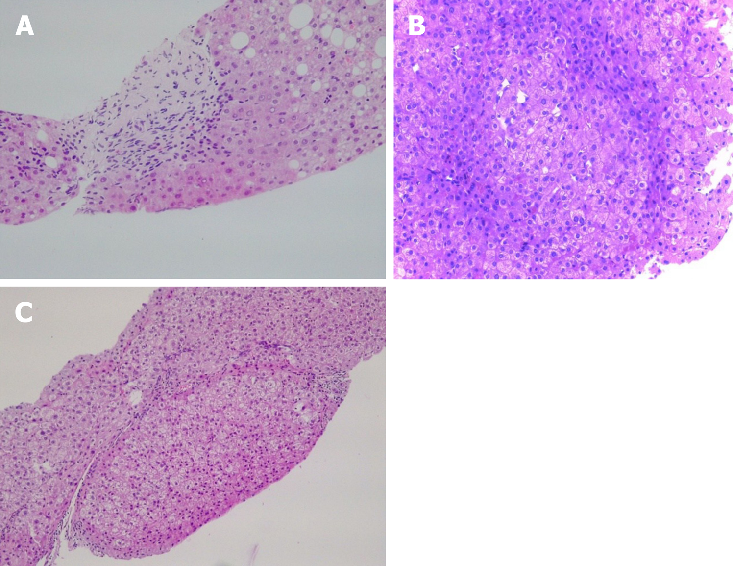
Figure 1 Pathological image.
A: Obliterative portal venopathy. Portal tract with round fibrous enlargement and vanishing of the portal vein radicle [Hematoxylin-eosin (HE) staining, 200×]; B: Liver biopsy showing nodular regenerative hyperplasia with vague nodularity of the parenchyma and compression of the adjacent hepatic (HE staining, 200×); C: Incomplete septal cirrhosis with delicate fibrous septa and no cirrhotic-type nodule formation is seen (HE staining, 200×).
- Citation: Nicoară-Farcău O, Rusu I, Stefănescu H, Tanțău M, Badea RI, Procopeț B. Diagnostic challenges in non-cirrhotic portal hypertension - porto sinusoidal vascular disease. World J Gastroenterol 2020; 26(22): 3000-3011
- URL: https://www.wjgnet.com/1007-9327/full/v26/i22/3000.htm
- DOI: https://dx.doi.org/10.3748/wjg.v26.i22.3000


