©The Author(s) 2019.
World J Gastroenterol. Feb 21, 2019; 25(7): 880-887
Published online Feb 21, 2019. doi: 10.3748/wjg.v25.i7.880
Published online Feb 21, 2019. doi: 10.3748/wjg.v25.i7.880
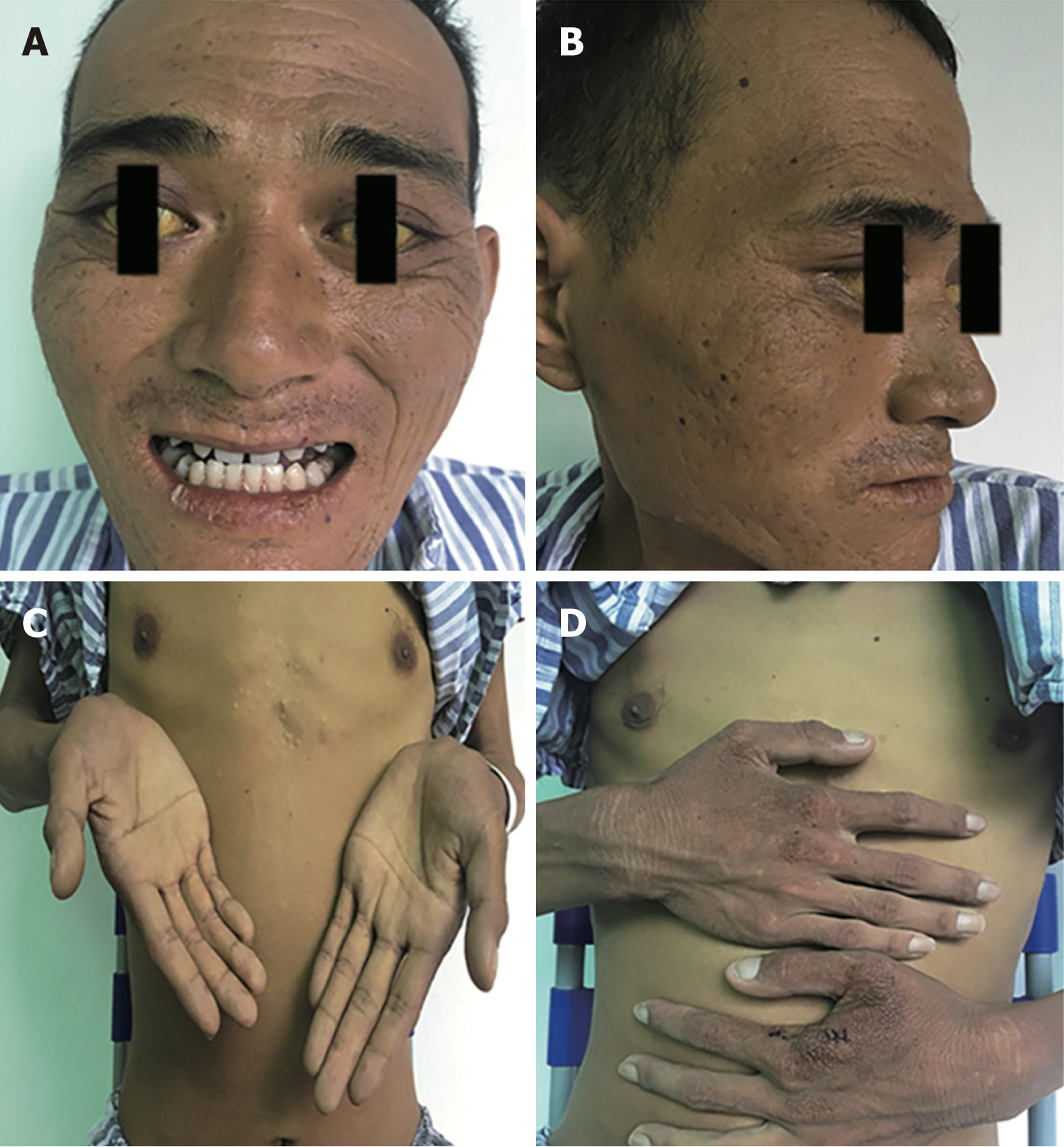
Figure 1 Clinical features.
Physical examination after admission showed severe yellowing of skin and sclera, rough and irregular skin on the arm, face and perioral area, and lichenification in some areas.
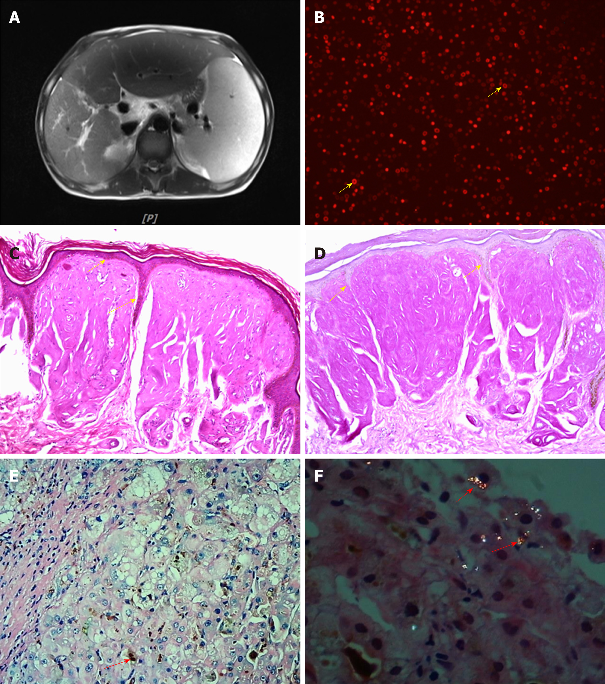
Figure 2 Accessory examinations.
A: Abdominal image of magnetic resonance imaging; B: Peripheral blood (×20); C: Hematoxylin and eosin (HE)-stained skin biopsy (×100); D: Periodic acid-Schiff-stained skin biopsy (×100); E: HE-stained liver biopsy under a light microscope; F: HE-stained liver biopsy under a polarized light microscope.
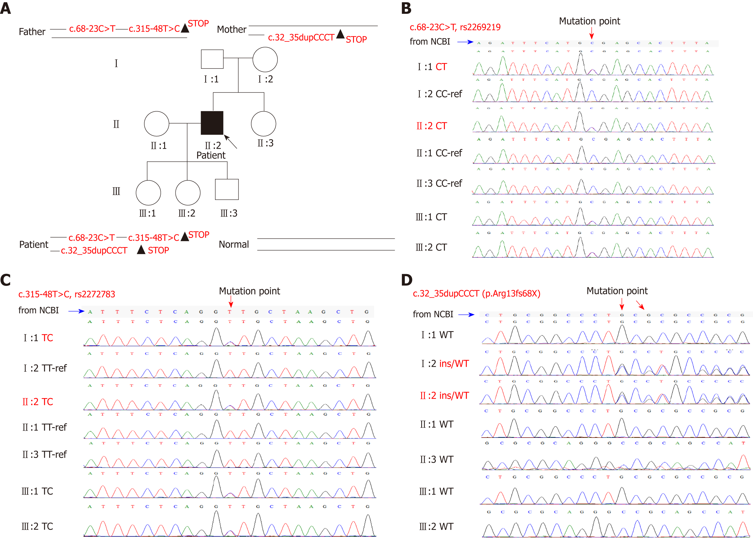
Figure 3 First generation sequencing.
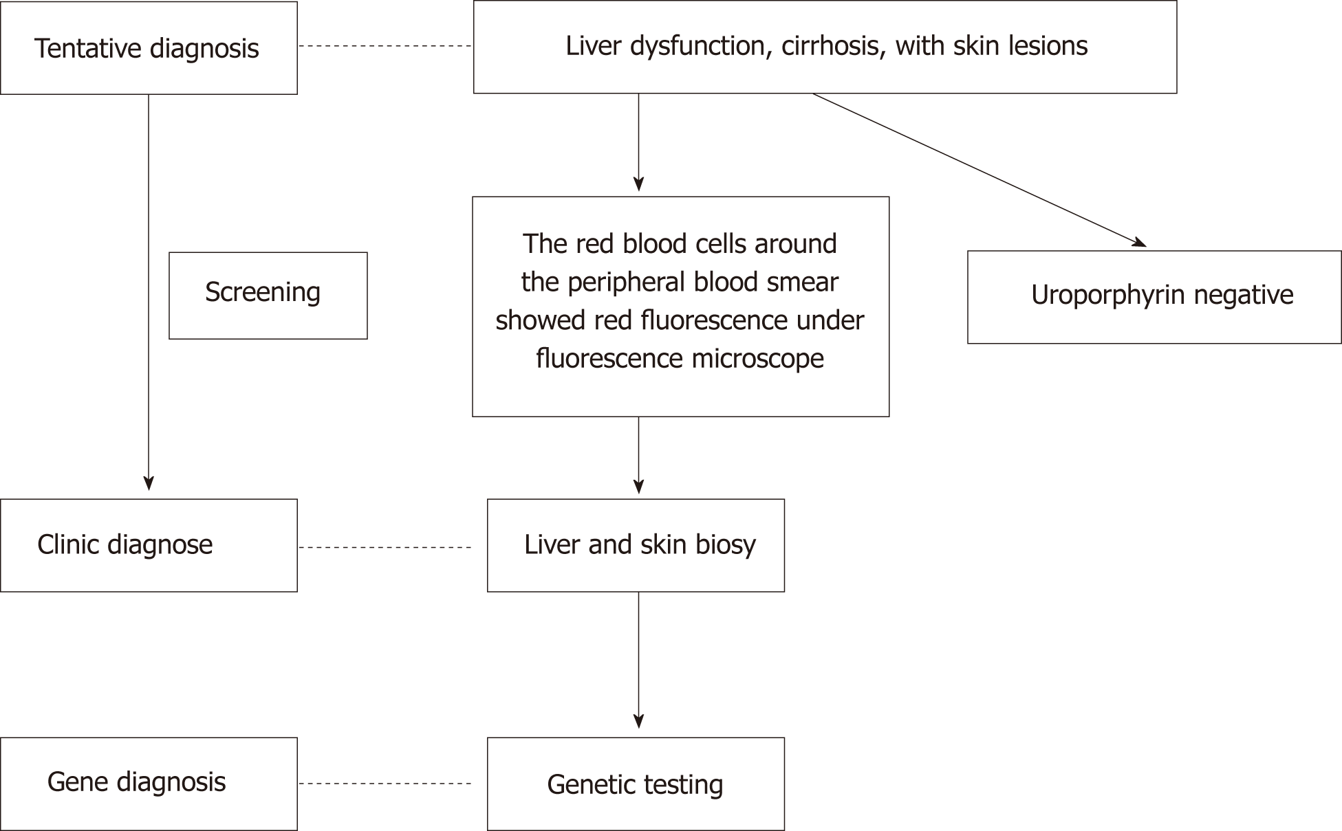
Figure 4 The diagnostic flow chart.
- Citation: Liu HM, Deng GH, Mao Q, Wang XH. Diagnosis of erythropoietic protoporphyria with severe liver injury: A case report. World J Gastroenterol 2019; 25(7): 880-887
- URL: https://www.wjgnet.com/1007-9327/full/v25/i7/880.htm
- DOI: https://dx.doi.org/10.3748/wjg.v25.i7.880


