©The Author(s) 2017.
World J Gastroenterol. Dec 7, 2017; 23(45): 8090-8096
Published online Dec 7, 2017. doi: 10.3748/wjg.v23.i45.8090
Published online Dec 7, 2017. doi: 10.3748/wjg.v23.i45.8090
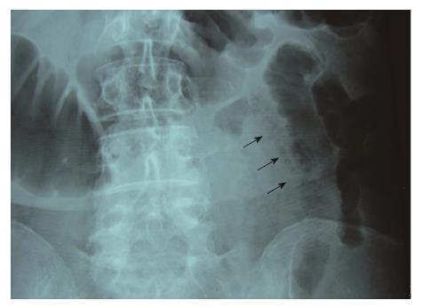
Figure 1 Plain abdominal X-ray depicts slightly dilated small bowel loop with pattern of intramural pearls of air (black arrows).
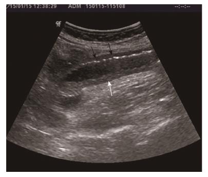
Figure 2 Sonographic features show dilated small bowel loop with absent peristalsis.
Also depicted are increased intraluminal secretions within the ischemic small bowel segment (white arrow), slight mural thickening and intramural gas (black arrows).
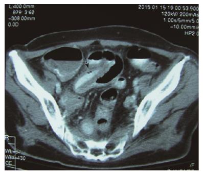
Figure 3 Axial contrast enhanced computed tomography demonstrates intestinal pneumatosis (long black arrow).
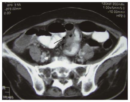
Figure 4 Axial contrast enhanced computed tomography demonstrates a homogeneous thickened soft tissue mass at the small bowel mesentery (long black arrows), as well as intestinal pneumatosis (small black arrows).
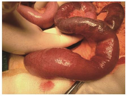
Figure 5 Intraoperative findings.
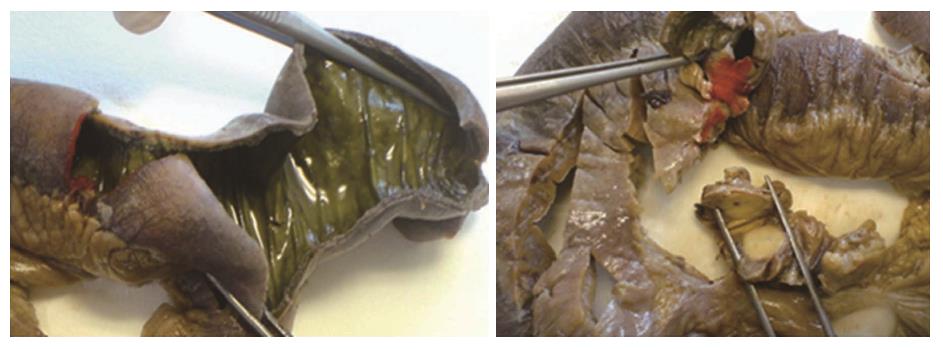
Figure 6 Gross pathology specimen of resected small bowel, showing ischemic bowel and enlarged mesenteric lymph nodes.
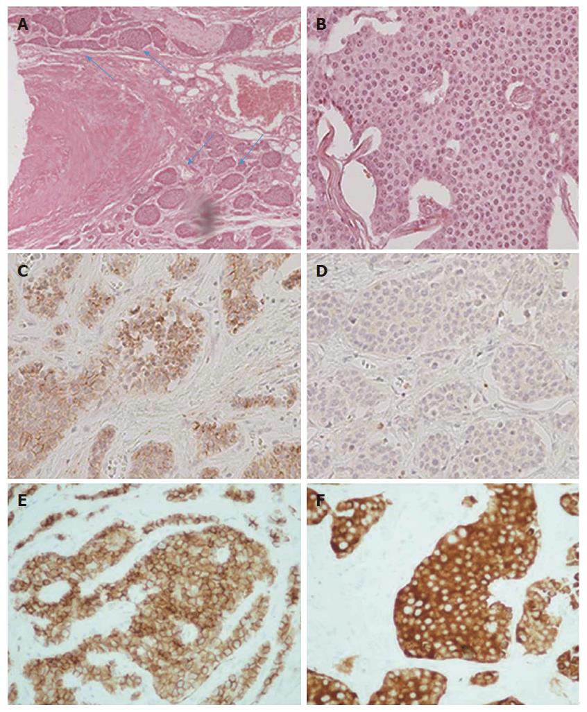
Figure 7 Histopathological findings.
A: Vascular and perineural invasion of tumor cells (blue arrows), medium magnification (100 ×); B: Tumor cells with characteristic nuclear appearance, high magnification (400 ×); C: Immunohistochemical staining reveals strong positivity for chromogranin A marker, high magnification (400 ×); D: Less than 1% of tumor cells reveal positivity for proliferative marker Ki-67, high magnification (400 ×); E: Immunohistochemical staining reveals strong positivity for CD56 marker, high magnification (400 ×); F: Immunohistochemical staining reveals strong positivity for synaptophysin marker, high magnification (400 ×).
- Citation: Mantzoros I, Savvala NA, Ioannidis O, Parpoudi S, Loutzidou L, Kyriakidou D, Cheva A, Intzos V, Tsalis K. Midgut neuroendocrine tumor presenting with acute intestinal ischemia. World J Gastroenterol 2017; 23(45): 8090-8096
- URL: https://www.wjgnet.com/1007-9327/full/v23/i45/8090.htm
- DOI: https://dx.doi.org/10.3748/wjg.v23.i45.8090


