©The Author(s) 2017.
World J Gastroenterol. Sep 14, 2017; 23(34): 6315-6320
Published online Sep 14, 2017. doi: 10.3748/wjg.v23.i34.6315
Published online Sep 14, 2017. doi: 10.3748/wjg.v23.i34.6315
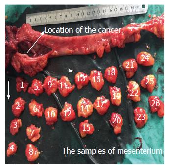
Figure 1 Samples of mesenterium from colorectal cancer patients.
Analysis of large cross sectional tissue samples of mesentery of colorectum from surgically resected specimens.
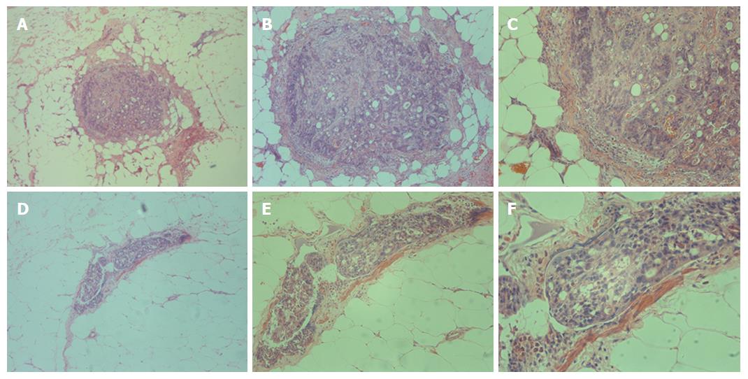
Figure 2 Detection of Metastasis V in colorectal cancer patients.
Isolated cancer cells were detected in the mesentery of colorectum by HE staining. A (× 10), B (× 20) and C (× 40) represent the same slide; D (× 10), E (× 20) and F (× 40) represent the same slide.
- Citation: Luo XL, Xie DX, Wu JX, Wu AD, Ge ZQ, Li HJ, Hu JB, Cao ZX, Gong JP. Detection of metastatic cancer cells in mesentery of colorectal cancer patients. World J Gastroenterol 2017; 23(34): 6315-6320
- URL: https://www.wjgnet.com/1007-9327/full/v23/i34/6315.htm
- DOI: https://dx.doi.org/10.3748/wjg.v23.i34.6315


