©The Author(s) 2017.
World J Gastroenterol. Aug 14, 2017; 23(30): 5619-5633
Published online Aug 14, 2017. doi: 10.3748/wjg.v23.i30.5619
Published online Aug 14, 2017. doi: 10.3748/wjg.v23.i30.5619
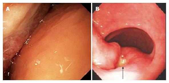
Figure 1 Findings at esophagogastroduodenoscopy in two patients with giant gastric lipomas.
A: Patient 1. Esophagogastroduodenoscopy (EGD) in a 63-year-old male who presented with melena and a hemoglobin decline to 6.2 g/dL that required transfusion of 2 units of packed erythrocytes, showing the distal body and antrum with a huge mass folded upon itself occupying most of the lumen and an 8 mm wide, nonbleeding, acute mucosal ulcer without stigmata of recent hemorrhage embedded deep in the valley (fold) between the right and left parts of the mass. The ulcer was attributed to friction from the opposing surface. The mass was 13-cm-wide, submucosal, yellowish, and covered by smooth mucosa except for focal ulceration, findings consistent with a gastric lipoma; B: Patient 2. EGD in a 78-year-old-woman, who presented with melena for 3 d, orthostatic dizziness, and a hemoglobin decline to 7.1 g/dL requiring transfusion of 2 units of packed erythrocytes, revealed an acute 1-cm-wide prepyloric ulcer (arrow) with a white exudate but without stigmata of recent hemorrhage between the right and left lobes of a large, well-demarcated, submucosal, mass covered by otherwise normal, superficial mucosa. This endoscopic photograph shows only a part of the mass.
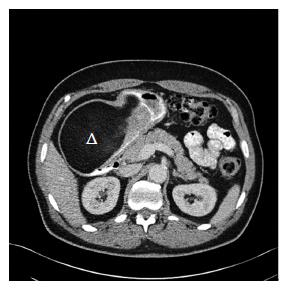
Figure 2 Abdominal computerized tomography findings in patient 1.
A 63-year-old male (patient 1) presented with acute melena and hemoglobin decline to 6.2 g/dL, and esophagogastroduodenoscopy revealed a huge, submucosal mass with a smooth overlying surface and exhibiting the pillow sign characteristic of a submucosal lipoma. Illustrated abdominal computerized tomography shows an approximately 13.4 cm × 8.2 cm × 8.4 cm mostly homogeneous, hypodense mass with a characteristic attenuation of fat (-90.2 Hounsfield units) extending from proximal gastric body through entire antrum. The normal-appearing very proximal stomach is filled with oral contrast without a mass, and leads to a very narrow, compressed, distal and dorsal, gastric channel containing oral contrast that passes into the duodenum. Triangle: antral giant gastric lipoma which has the characteristic hypodense attenuation of fat.
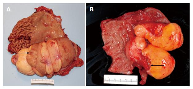
Figure 3 Gross pathologic findings in gastrectomy specimens in two patients with giant gastric lipomas.
A: Patient 1. Patient 1 presented with acute melena and hemoglobin decline and had an ulcer detected at esophagogastroduodenoscopy (EGD) within a huge, lipomatous gastric mass. Gross pathologic view of the gastrectomy specimen after it is opened to expose the luminal surface shows a well-circumscribed, lobulated, 14.5 cm × 9.0 cm × 7.5 cm lipomatous mass extending from the gastric body (left) to antrum (right). A small ulcer (round depression, arrow) is present on the mucosa overlying the lipomatous mass. Normal gastric rugae are present above the mass on the upper left, but have been effaced on the upper right, likely because of chronic compression/pressure from the giant lipomatous mass located below (on the contralateral gastric wall before opening the stomach). Vertical incisions show a homogeneous yellow-tan cut surface, indicative of a lipomatous tumor; B: Patient 2. Patient 2 presented with melena for 3 d, orthostatic dizziness, and a hemoglobin decline to 7.1 g/dL requiring transfusion of 2 units of packed erythrocytes and had at EGD a large, yellowish, smooth, well-circumscribed antral mass. Gross pathologic view of distal gastrectomy specimen after it is opened to expose the luminal surface shows normal gastric antral tissue at left and a lobulated, well-circumscribed, yellow-tan, 9.0 cm × 6.0 cm × 3.5 cm lipomatous tumor at right, with a deep, clean-based, ulcer (arrow) on the mucosa overlying the mass.
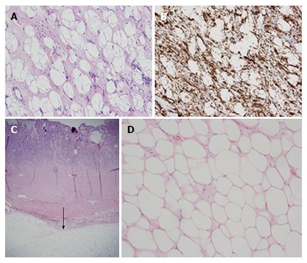
Figure 4 Histopathologic findings in gastrectomy specimens in two patients with giant gastric lipomas.
A: Patient 1-standard histochemistry. Medium power photomicrograph of a hematoxylin and eosin stain of a tissue section from the resected gastric mass in patient 1 reveals large adipocytes filled with clear, homogeneous, cytoplasm and tiny, compressed, peripheral nuclei. No lipoblasts are detected. Note the spindle-shaped stroma surrounding the adipocytes, findings consistent with spindle cell lipoma, as proven by immunohistochemistry (B); B: Patient 1-immunohistochemistry. Medium power photomicrograph of immunohistochemistry, using an antibody to CD34, reveals within tumor in patient 1 extensive staining in a spindly pattern of the stroma surrounding characteristically clear adipocytes, a characteristic staining pattern for spindle-shaped lipoma; C: Patient 2-standard histochemistry-low power. Low power photomicrograph of a hematoxylin and eosin stain of a tissue section from resected gastric mass in patient 2 reveals a well-circumscribed, submucosal layer composed of adipocytes with clear cytoplasm (arrow) and scant loose, myxoid stroma; D: Patient 2-standard histochemistry-medium power. Medium power photomicrograph of a hematoxylin and eosin stain of a tissue section from resected gastric mass in patient 2 reveals sheets of large, adipocytes filled with clear, homogeneous, cytoplasm and tiny, compressed, peripheral nuclei, with scant loose, myxoid stroma. No lipoblasts are detected. These histologic findings are characteristic of lipomas.
- Citation: Cappell MS, Stevens CE, Amin M. Systematic review of giant gastric lipomas reported since 1980 and report of two new cases in a review of 117110 esophagogastroduodenoscopies. World J Gastroenterol 2017; 23(30): 5619-5633
- URL: https://www.wjgnet.com/1007-9327/full/v23/i30/5619.htm
- DOI: https://dx.doi.org/10.3748/wjg.v23.i30.5619


