Copyright
©The Author(s) 2017.
World J Gastroenterol. Jan 7, 2017; 23(1): 185-190
Published online Jan 7, 2017. doi: 10.3748/wjg.v23.i1.185
Published online Jan 7, 2017. doi: 10.3748/wjg.v23.i1.185
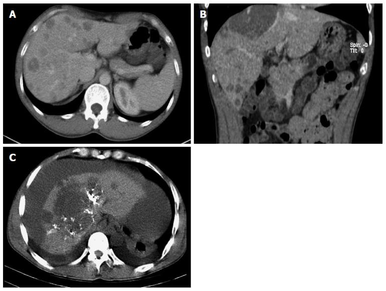
Figure 1 Computer tomography.
A: Venous phase image revealed multiple low-density intrahepatic nodules in both lobes, and part of nodules showed inhomogeneous enhancement; B: The maximum lesion was located in the right lobe under the hepatic capsule; C: Computer tomography scan after transcatheter arterial embolization revealed massive hematocoelia.
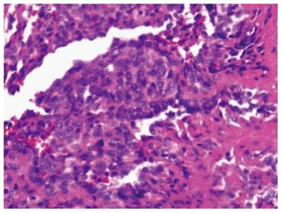
Figure 2 Histopathologic findings of hepatic epithelioid hemangioendothelioma.
Carcinoma cells appeared nest-like or papillary around the blood vessels, the nuclei were round or ovoid (hematoxylin-eosin staining; magnification × 200).
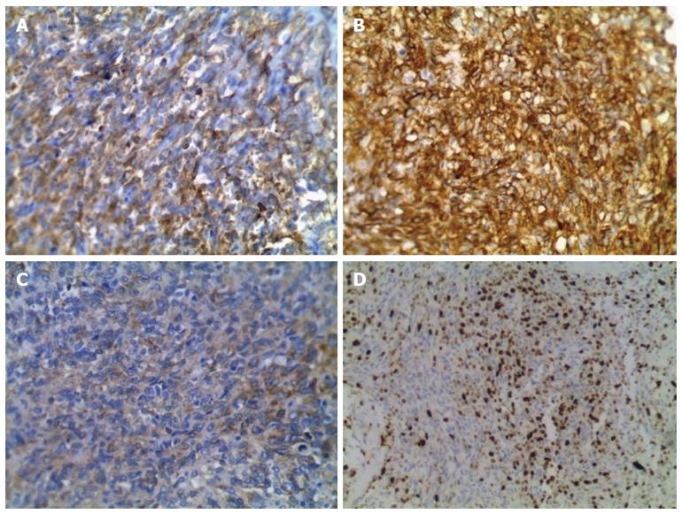
Figure 3 Immunohistochemical examination of hepatic epithelioid hemangioendothelioma.
A: Specimen stained positive for CD31 (magnification × 200); B: Specimen stained positive for CD34 (magnification × 200); C: Specimen stained positive for FVIII (magnification × 200); D: Ki-67 proliferation index is less than 40% (magnification × 100).
- Citation: Yang JW, Li Y, Xie K, Dong W, Cao XT, Xiao WD. Spontaneous rupture of hepatic epithelioid hemangioendothelioma: A case report. World J Gastroenterol 2017; 23(1): 185-190
- URL: https://www.wjgnet.com/1007-9327/full/v23/i1/185.htm
- DOI: https://dx.doi.org/10.3748/wjg.v23.i1.185


