Copyright
©The Author(s) 2017.
World J Gastroenterol. Jan 7, 2017; 23(1): 173-177
Published online Jan 7, 2017. doi: 10.3748/wjg.v23.i1.173
Published online Jan 7, 2017. doi: 10.3748/wjg.v23.i1.173
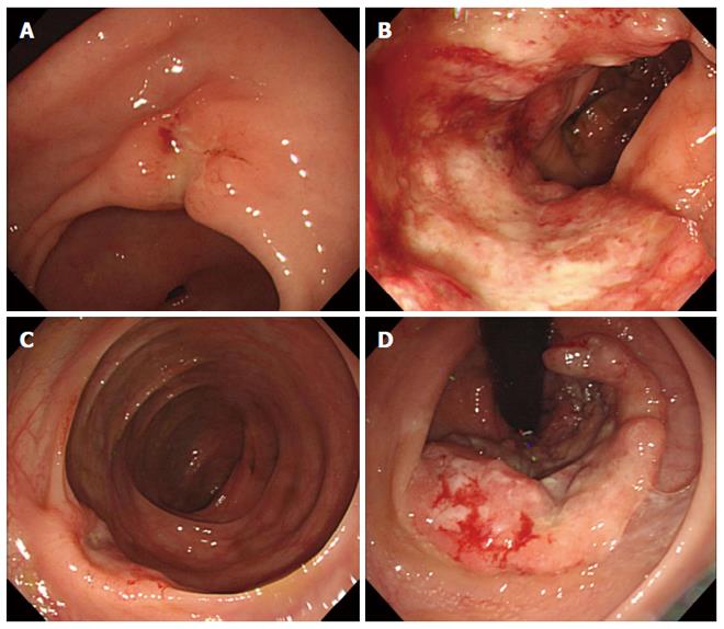
Figure 1 Endoscopic findings.
A: Early gastric cancer type IIc lesion at antrum, posterior wall of stomach; B: Ulcerative mass at proximal ascending colon, diagnosed with adenocarcinoma; C: Concave mass at transverse colon, diagnosed with adenocarcinoma; D: Ulcerative rectal mass near anus, diagnosed with adenocarcinoma pathologically.
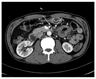
Figure 2 Computed tomography finding.
Focal irregular wall thickening at proximal jejunum with lymphadenopathy, suggesting adenocarcinoma.
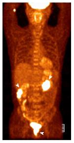
Figure 3 Positron emission tomography/computed tomography findings.
Revealed abnormal increased fluorodeoxy glucose (FDG) uptakes in ascending colon, rectum and jejunum but no definite abnormal FDG uptake along the gastric wall and transverse colon was noted. Diffuse increased FDG uptake was found at both thyroid glands, which allowed for the diagnosis of thyroiditis.
- Citation: Kim SH, Park BS, Kim HS, Kim JH. Synchronous quintuple primary gastrointestinal tract malignancies: Case report. World J Gastroenterol 2017; 23(1): 173-177
- URL: https://www.wjgnet.com/1007-9327/full/v23/i1/173.htm
- DOI: https://dx.doi.org/10.3748/wjg.v23.i1.173


