Copyright
©The Author(s) 2016.
World J Gastroenterol. Nov 21, 2016; 22(43): 9623-9630
Published online Nov 21, 2016. doi: 10.3748/wjg.v22.i43.9623
Published online Nov 21, 2016. doi: 10.3748/wjg.v22.i43.9623
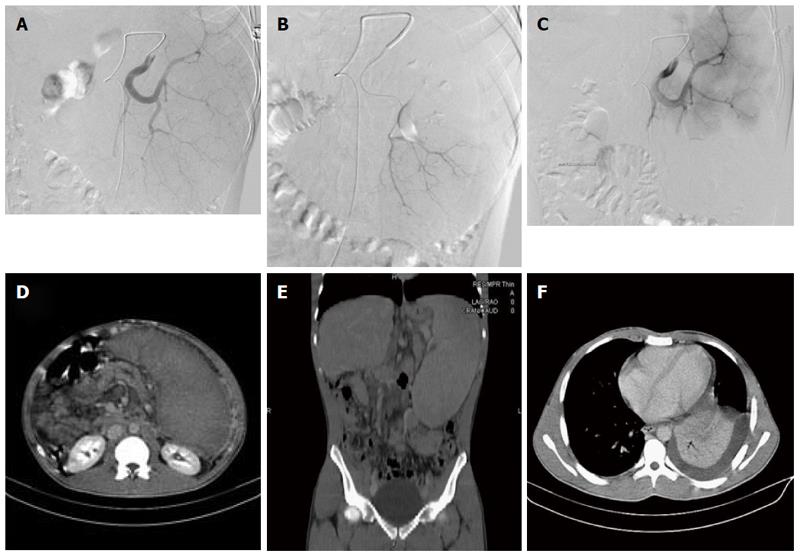
Figure 1 Radiographic images of patient No.
10. A: Selective splenic arteriography showing the multiple splenic vessels supplying the enlarged spleen; B: Super selective catheterization of the inferior branches supplying the lower pole of the spleen; C: Post embolization images of the splenic artery, with no enhancement observed in the lower pole due to the embolized inferior lobe branch; D: Post procedural 1-mo follow-up with axial CT images, with no enhancement observed in the lower embolized portion of the spleen; E: Pre-procedural coronal reformatted CT images showing the enlarged spleen; F: Post-procedural axial CT image showing left pleural effusion accompanying lower lobe atelectasis.
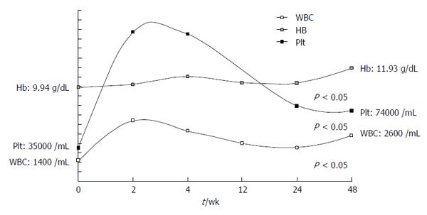
Figure 2 Blood counts during follow-up post partial splenic embolization.
- Citation: Ozturk O, Eldem G, Peynircioglu B, Kav T, Görmez A, Cil BE, Balkancı F, Sokmensuer C, Bayraktar Y. Outcomes of partial splenic embolization in patients with massive splenomegaly due to idiopathic portal hypertension. World J Gastroenterol 2016; 22(43): 9623-9630
- URL: https://www.wjgnet.com/1007-9327/full/v22/i43/9623.htm
- DOI: https://dx.doi.org/10.3748/wjg.v22.i43.9623


