Copyright
©The Author(s) 2016.
World J Gastroenterol. Nov 14, 2016; 22(42): 9411-9418
Published online Nov 14, 2016. doi: 10.3748/wjg.v22.i42.9411
Published online Nov 14, 2016. doi: 10.3748/wjg.v22.i42.9411
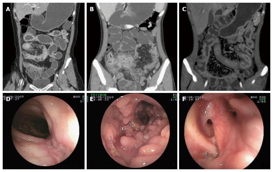
Figure 1 Endoscopic and computed tomographic enterography features in Crohn’s disease.
A: Coronary reconstructed CTE reflected stricture with proximal dilation and comb sign in ileum in ileum; B: Phelgmon in distal ileum; C: Stretching and densifying of distal mesenteric artery so-called comb sign in ileum; D: DBE found longitudinal ulcer in distal ileum; E: Colonoscopy detected Pseudo-polyps formation in ascending colon; F: Perianal involvement in CD.
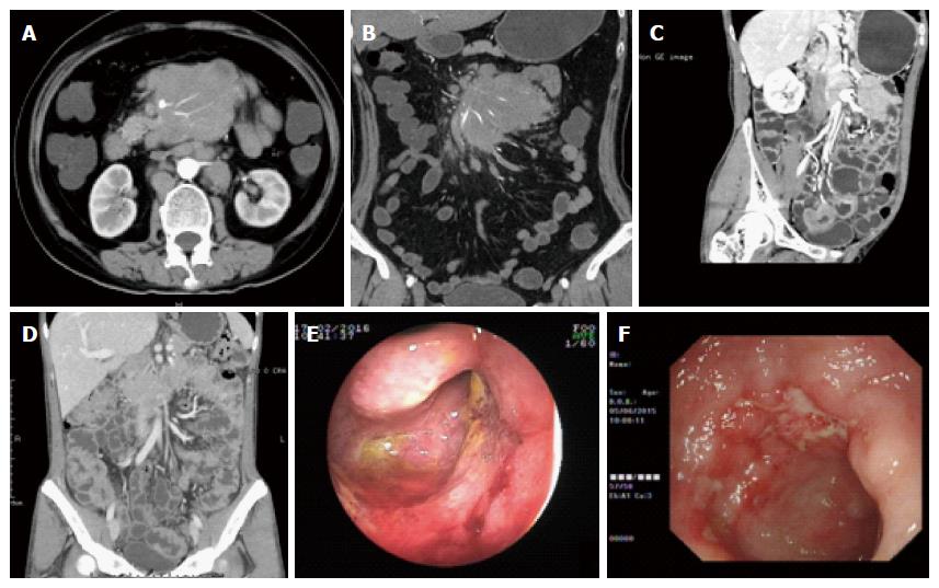
Figure 2 Endoscopic and computed tomographic enterography features in primary intestinal lymphoma.
A and B: Axial and coronary reconstructed CTE sections showed mass of sandwich sign in mensentery area; C: Coronary reconstructed CTE reflected aneurysmal dilation in pelvic intestine; D: Coronary reconstructed CTE displayed circular thickening of bowel wall without stricture in ileocecal region; E: DBE showed intraluminal proliferative mass in proximal ileum; F: Colonoscopy revealed irregular ulcer in ileocecal region.
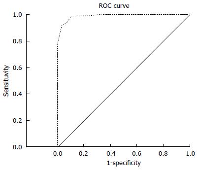
Figure 3 Receiver operating characteristic curve of differentiating model between Crohn’s disease and primary intestinal lymphoma (area under the ROC curve = 0.
989). ROC: Receiver operating characteristic.
- Citation: Zhang TY, Lin Y, Fan R, Hu SR, Cheng MM, Zhang MC, Hong LW, Zhou XL, Wang ZT, Zhong J. Potential model for differential diagnosis between Crohn's disease and primary intestinal lymphoma. World J Gastroenterol 2016; 22(42): 9411-9418
- URL: https://www.wjgnet.com/1007-9327/full/v22/i42/9411.htm
- DOI: https://dx.doi.org/10.3748/wjg.v22.i42.9411


