©The Author(s) 2016.
World J Gastroenterol. Nov 7, 2016; 22(41): 9229-9234
Published online Nov 7, 2016. doi: 10.3748/wjg.v22.i41.9229
Published online Nov 7, 2016. doi: 10.3748/wjg.v22.i41.9229
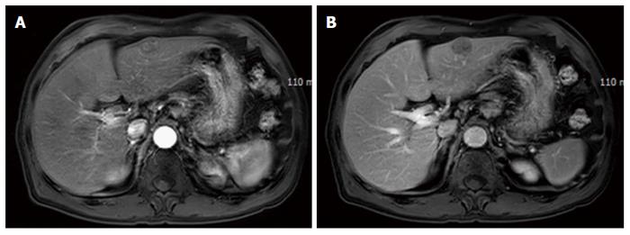
Figure 1 Magnetic resonance imaging of the liver.
A 2.2 cm × 2.2 cm sized lobular contoured mass was found on segment 3. It showed mild enhancement in the arterial phase (A) and a washed-out pattern in the portal phase (B).
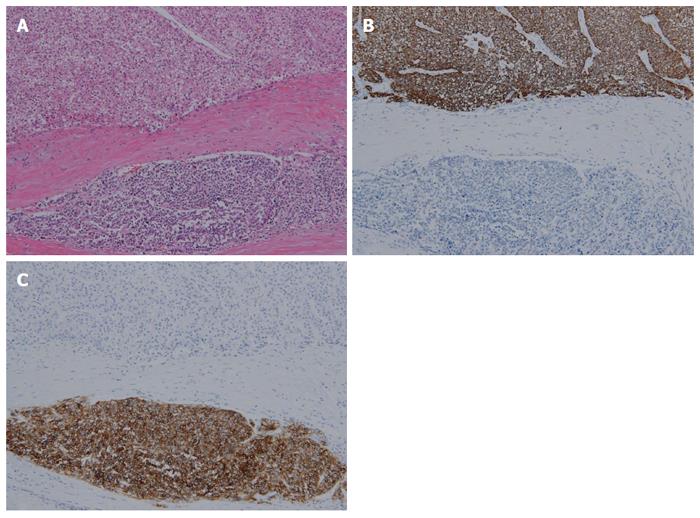
Figure 2 Microscopic findings.
A: Moderately differentiated hepatocellular carcinoma (HCC) is found in the upper portion. The malignant cells show a clear and rich cytoplasm. It is separated from neuroendocrine carcinoma (NEC) by a fibrous band. Poorly differentiated NEC is found in the lower portion. The cytoplasm is barely seen, and the N/C (nucleus/cytoplasm) is very high (hematoxylin eosin staining, magnification × 100); B: Immunohistochemical staining of hepatocyte paraffin-1 is positive in the upper HCC portion (magnification × 100); C: Immunohistochemical staining of CD56 is positive in the lower NEC portion (magnification × 100).
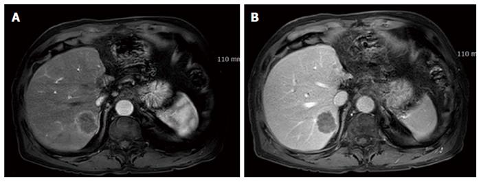
Figure 3 Magnetic resonance imaging of the liver after 6 mo.
Five nodules were detected in the right lobe. The biggest was 3.3 cm. The nodules showed rim enhancement on the arterial phase (A) and low density on the portal phase (B).
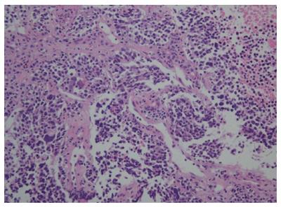
Figure 4 Microscopic findings of fine-needle biopsy.
The size of malignant cells is variable, and the shape is very bizarre. They are clustered, forming nests, and the mitosis rate is above 20 per 10 HPF (hematoxylin eosin, magnification × 100).
- Citation: Choi GH, Ann SY, Lee SI, Kim SB, Song IH. Collision tumor of hepatocellular carcinoma and neuroendocrine carcinoma involving the liver: Case report and review of the literature. World J Gastroenterol 2016; 22(41): 9229-9234
- URL: https://www.wjgnet.com/1007-9327/full/v22/i41/9229.htm
- DOI: https://dx.doi.org/10.3748/wjg.v22.i41.9229


