Copyright
©The Author(s) 2016.
World J Gastroenterol. Sep 14, 2016; 22(34): 7851-7856
Published online Sep 14, 2016. doi: 10.3748/wjg.v22.i34.7851
Published online Sep 14, 2016. doi: 10.3748/wjg.v22.i34.7851
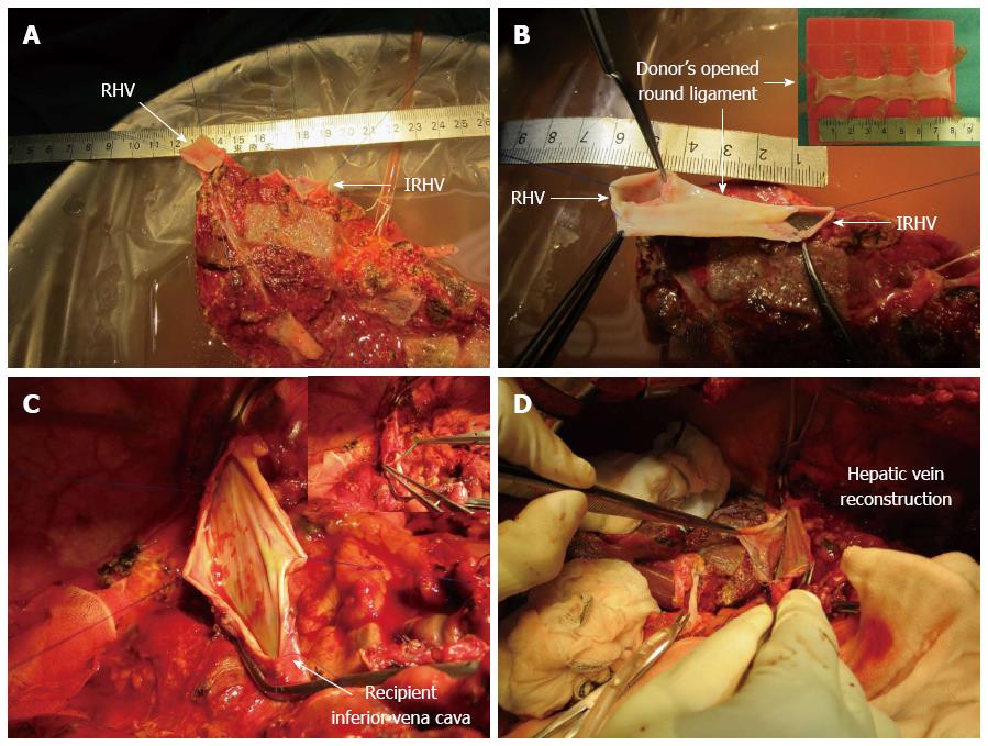
Figure 1 The graft had two hepatic veins.
Including the right hepatic vein (RHV; 15 mm) and the inferior RHV (IRHV; 20 mm) (A). The graft RHV and IRHV were formed into a single orifice using the donor’s opened round ligament (60 mm × 20 mm) as a venous patch graft during bench surgery (B). It was then anastomosed end-to-side with the recipient inferior vena cava (C and D).
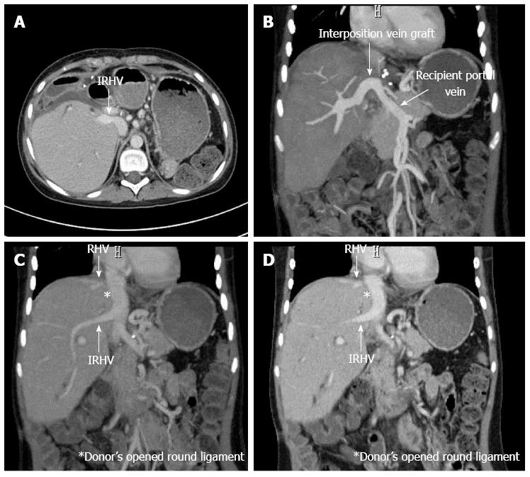
Figure 2 Abdominal enhanced computed tomography on post-operative day 82.
The radiological patency of the donor’s opened round ligament and the hepatic vein was confirmed (A, B, C, D). RHV: Right hepatic vein; IRHV: Inferior right hepatic vein.
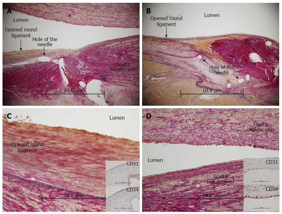
Figure 3 Pathology of the hepatic venous anastomotic site on autopsy.
No stenosis or thrombus was identified (A and B). On pathology, there was adequate patency and continuity between the recipient’s hepatic vein and the donor’s opened round ligament. In addition, the stains for CD31 and CD34 on the inner membrane of the opened round ligament were positive, a finding also observed in the graft hepatic vein (C and D).
- Citation: Sanada Y, Sakuma Y, Sasanuma H, Miki A, Katano T, Hirata Y, Okada N, Yamada N, Ihara Y, Urahashi T, Sata N, Yasuda Y, Mizuta K. Immunohistochemical evaluation for outflow reconstruction using opened round ligament in living donor right posterior sector graft liver transplantation: A case report. World J Gastroenterol 2016; 22(34): 7851-7856
- URL: https://www.wjgnet.com/1007-9327/full/v22/i34/7851.htm
- DOI: https://dx.doi.org/10.3748/wjg.v22.i34.7851


