©The Author(s) 2016.
World J Gastroenterol. Jun 21, 2016; 22(23): 5342-5352
Published online Jun 21, 2016. doi: 10.3748/wjg.v22.i23.5342
Published online Jun 21, 2016. doi: 10.3748/wjg.v22.i23.5342
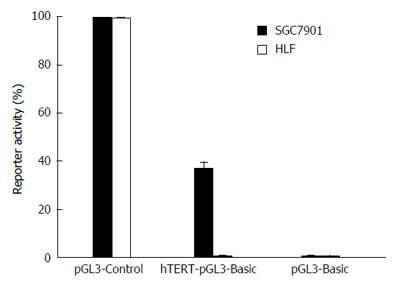
Figure 1 Human telomerase reverse transcriptase promoter activities in cancerous and normal cells.
EC9706 and HLF cells were transfected with hTERT reporter vector (hTERT-pGL3 basic). pGL3-Control and pGL3 basic were transfected as positive and negative controls, respectively. hTERT: Human telomerase reverse transcriptase.
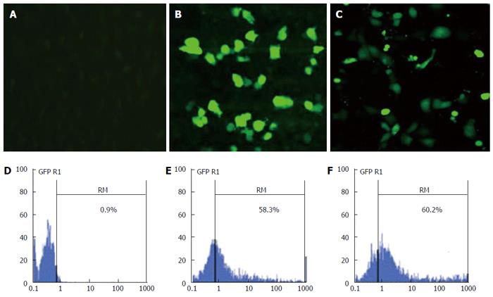
Figure 2 Transfection efficiency of calcium phosphate nanoparticles.
A-C: Images of transfected EC9706 cells acquired by fluorescence microscope at magnification × 200 after 48 h of transfection. Negative control (A), CPNP-GFP complex (B), Liposome/GFP complex (C); D-F: Qualitative analysis of transfection efficiency by flow cytometric assay. Negative control (D), CPNP-GFP complex (E), Liposome/GFP complex (F). CPNP: Calcium phosphate nanoparticles; GFP: Green fluorescent protein.
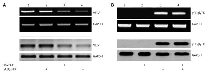
Figure 3 Changes of vascular endothelial growth factor and yCDglyTK expression in established stable cell lines.
A: Representative VEGF mRNA and protein expression were analyzed by RT-PCR (top panel) and western blot (bottom panel), respectively. GAPDH was used as an internal control. Lane 1, EC9706/null; lane 2, EC9706/yCDglyTK; lane 3, EC9706/shVEGF; lane 4, EC9706/shVEGF-yCDglyTK; B: Representative yCDglyTK mRNA and protein expression were analyzed by semiquantitative RT-PCR (top panel) and western blot (bottom panel), respectively. GAPDH was used as an internal control. Lane 1, EC9706/null; lane 2, EC9706/shVEGF; lane 3, EC9706/yCDglyTK; lane 4, EC9706/shVEGF-yCDglyTK. GAPDH: Glyceraldehyde-3-phosphate dehydrogenase; RT-PCR: Reverse transcription polymerase chain reaction; VEGF: Vascular endothelial growth factor.
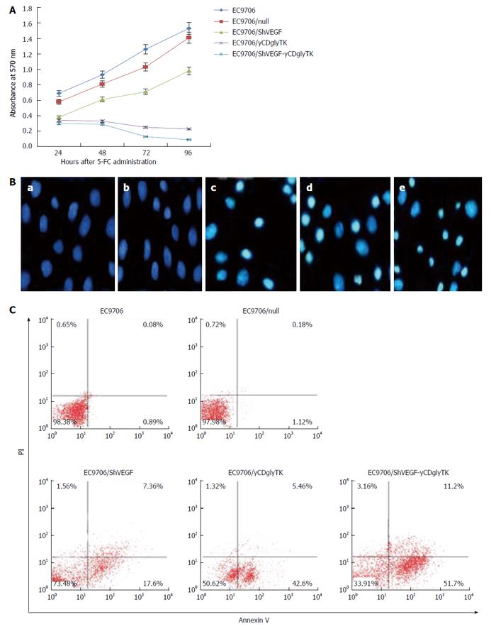
Figure 4 Effects of shVEGF-yCDglyTK/5-FC system on EC9706 cells.
A: Cell viability of parental and stable EC9706 cells were determined with MTT assay at various time points after 5-FC treatment. The results shown are representative of three independent experiments; B: Representative images of Hoechst 33258-stained nuclei at magnification × 200. Apoptotic nuclei are condensed or fragmented; parental EC9706 cells (a); EC9706/null (b); EC9706/shVEGF (c); EC9706/yCDglyTK (d); EC9706/shVEGF-yCDglyTK (e); C: Representative dot plots of flow cytometry analysis. The numbers represent the percentage (%) of cells. Upper right quadrant, late apoptosis; lower right quadrant, early apoptosis; lower left, live cells. 5-FC: 5-fluorocytosine.
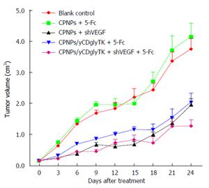
Figure 5 shVEGF-yCDglyTK/5-FC system inhibited tumor growth in the EC9706 xenograft model.
Twenty-five BALB/C nude mice bearing EC9706 xenografts were randomized into five groups. The CPNPs/null, CPNPs/shVEGF, CPNPs/yCDglyTK or CPNPs/shVEGF-yCDglyTK complexes were delivered by intratumoral injection every other day, and the injection was repeated three times in total. 5-FC (500 mg/kg) was administered daily for 14 consecutive days. Tumors were measured every 3 d. All of the mice were scarified at day 36 after inoculation.
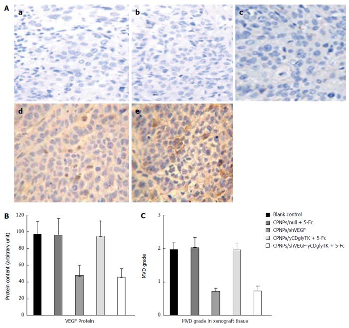
Figure 6 Immunohistochemistry analysis for yCDglyTK and vascular endothelial growth factor in EC9706 xenograft sections.
A: Histological expression and distribution of yCDglyTK at magnification × 200; no-treatment control group (a); CPNPs/null + 5-FC (b); CPNPs/shVEGFX (c); CPNPs/yCDglyTK+5-FC (d); CPNPs/shVEGF-yCDglyTK +5-FC (e); B: Integrated optical density (IOD) values of VEGF expression in EC9706 xenografts. Anti-VEGF antibody was used for immunohistochemistry assay; C: Quantification of angiogenesis by microvessel counts (MVC) in EC9706 xenografts. Anti-CD34 was used for microvessel staining. CPNPs: Calcium phosphate nanoparticles; VEGF: Vascular endothelial growth factor; 5-FC: 5-fluorocytosine.
- Citation: Liu T, Wu HJ, Liang Y, Liang XJ, Huang HC, Zhao YZ, Liao QC, Chen YQ, Leng AM, Yuan WJ, Zhang GY, Peng J, Chen YH. Tumor-specific expression of shVEGF and suicide gene as a novel strategy for esophageal cancer therapy. World J Gastroenterol 2016; 22(23): 5342-5352
- URL: https://www.wjgnet.com/1007-9327/full/v22/i23/5342.htm
- DOI: https://dx.doi.org/10.3748/wjg.v22.i23.5342


