©The Author(s) 2016.
World J Gastroenterol. Apr 21, 2016; 22(15): 4066-4070
Published online Apr 21, 2016. doi: 10.3748/wjg.v22.i15.4066
Published online Apr 21, 2016. doi: 10.3748/wjg.v22.i15.4066
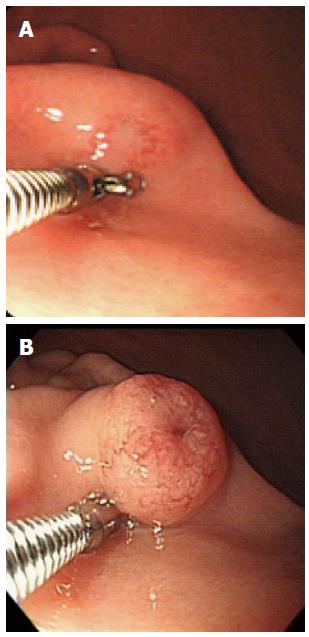
Figure 1 Gastric subepithelial tumor on the greater curvature side of the lower body.
A: 7 years ago, gastric subepithelial tumor (SET) with hypervascular mucosal change was shown. When pushed the gastric SET by cold biopsy forcep, mucous secretion flowed from the lesion; B: At admission, gastric SET had shown the growth of size. Top of the gastric SET was shown slightly depressed, irregular and hypervascular change.
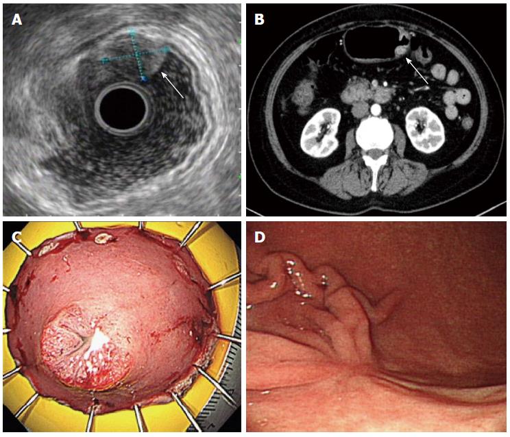
Figure 2 Colonoscopic examinations.
A: Endoscopic ultrasonography (EUS) demonstrated a 13.2 mm × 11.2 mm-sized heterogeneous hypoechoic tumor (white arrow) in the submucosal layer of the gastric wall; B: Abdominal contrast-enhanced computed tomography (CT) demonstrated a 1.5 cm-sized, oval-shaped enhancing mass (white arrow) on the GC side of the lower body; C: ESD specimen measuring 5.0 cm × 3.0 cm showed a 1.5 cm-sized, well-circumscribed polypoid lesion; D: Follow-up endoscopic examination demonstrated a post ESD scar with converging fold on the GC side of the lower body.
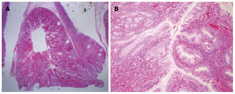
Figure 3 Microscopy of the endoscopic submucosal dissection specimen.
A: Well-circumscribed inverted growth pattern into the submucosal layer was shown (HE staining, × 10); B: Endophytic proliferation of hyperplastic columnar cells and connected with inflamed surface epithelium was shown. No architectural or cytological atypia was found (HE staining, × 100).
- Citation: Yun JT, Lee SW, Kim DP, Choi SH, Kim SH, Park JK, Jang SH, Park YJ, Sung YG, Sul HJ. Gastric inverted hyperplastic polyp: A rare cause of iron deficiency anemia. World J Gastroenterol 2016; 22(15): 4066-4070
- URL: https://www.wjgnet.com/1007-9327/full/v22/i15/4066.htm
- DOI: https://dx.doi.org/10.3748/wjg.v22.i15.4066


