Copyright
©The Author(s) 2016.
World J Gastroenterol. Mar 28, 2016; 22(12): 3496-3501
Published online Mar 28, 2016. doi: 10.3748/wjg.v22.i12.3496
Published online Mar 28, 2016. doi: 10.3748/wjg.v22.i12.3496
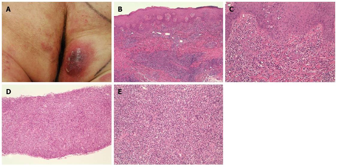
Figure 1 Macroscopic aspect of cutaneous lesions (A-C) and pancreatic lesion (D and E).
Hematoxilyn and eosin staining, magnification × 4 (B), × 10 (D), × 20 (C and E).
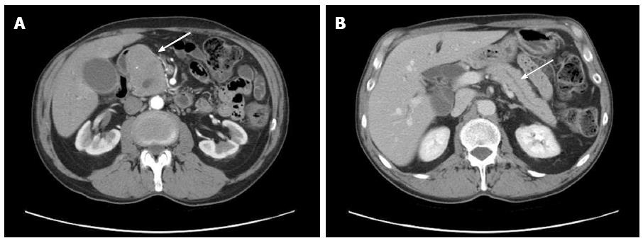
Figure 2 Abdominal computed tomography with contrast agent.
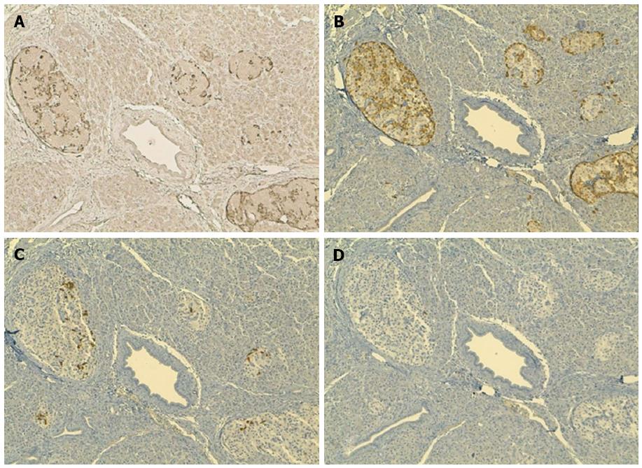
Figure 3 Immunohistochemical staining of pancreatic archival tissue sections.
A: CCL27; B: Glucagon; C: Somatostati; D: Pancreatic polypeptide. A-D: Magnifications × 10.
- Citation: Ceriolo P, Fausti V, Cinotti E, Bonadio S, Raffaghello L, Bianchi G, Orcioni GF, Fiocca R, Rongioletti F, Pistoia V, Borgonovo G. Pancreatic metastasis from mycosis fungoides mimicking primary pancreatic tumor. World J Gastroenterol 2016; 22(12): 3496-3501
- URL: https://www.wjgnet.com/1007-9327/full/v22/i12/3496.htm
- DOI: https://dx.doi.org/10.3748/wjg.v22.i12.3496


