©The Author(s) 2016.
World J Gastroenterol. Mar 21, 2016; 22(11): 3105-3116
Published online Mar 21, 2016. doi: 10.3748/wjg.v22.i11.3105
Published online Mar 21, 2016. doi: 10.3748/wjg.v22.i11.3105
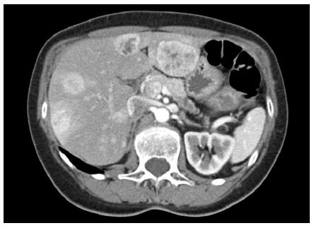
Figure 1 Computed tomography abdomen and pelvis with contrast showing multiple enhancing liver lesions and a probable primary tumor in the pancreatic body.
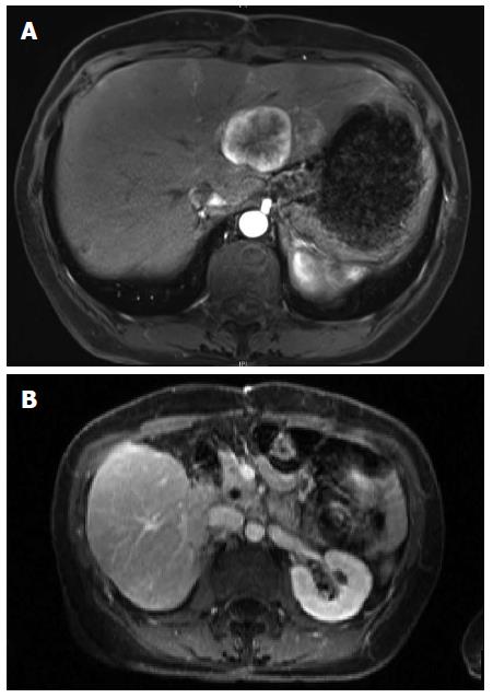
Figure 2 Magnetic resonance imaging.
A: T1 weighted magnetic resonance imaging showing persistent left lobe lesion after treatment of right sided lesions with radioembolization; B: Post-operative magnetic resonance imaging showing liver remnant and pancreatic head with superior mesenteric vein after left hepatectomy and distal pancreatectomy.
- Citation: Folkert IW, Hernandez P, Roses RE. Multidisciplinary management of nonfunctional neuroendocrine tumor of the pancreas. World J Gastroenterol 2016; 22(11): 3105-3116
- URL: https://www.wjgnet.com/1007-9327/full/v22/i11/3105.htm
- DOI: https://dx.doi.org/10.3748/wjg.v22.i11.3105


