©The Author(s) 2015.
World J Gastroenterol. Sep 28, 2015; 21(36): 10418-10426
Published online Sep 28, 2015. doi: 10.3748/wjg.v21.i36.10418
Published online Sep 28, 2015. doi: 10.3748/wjg.v21.i36.10418
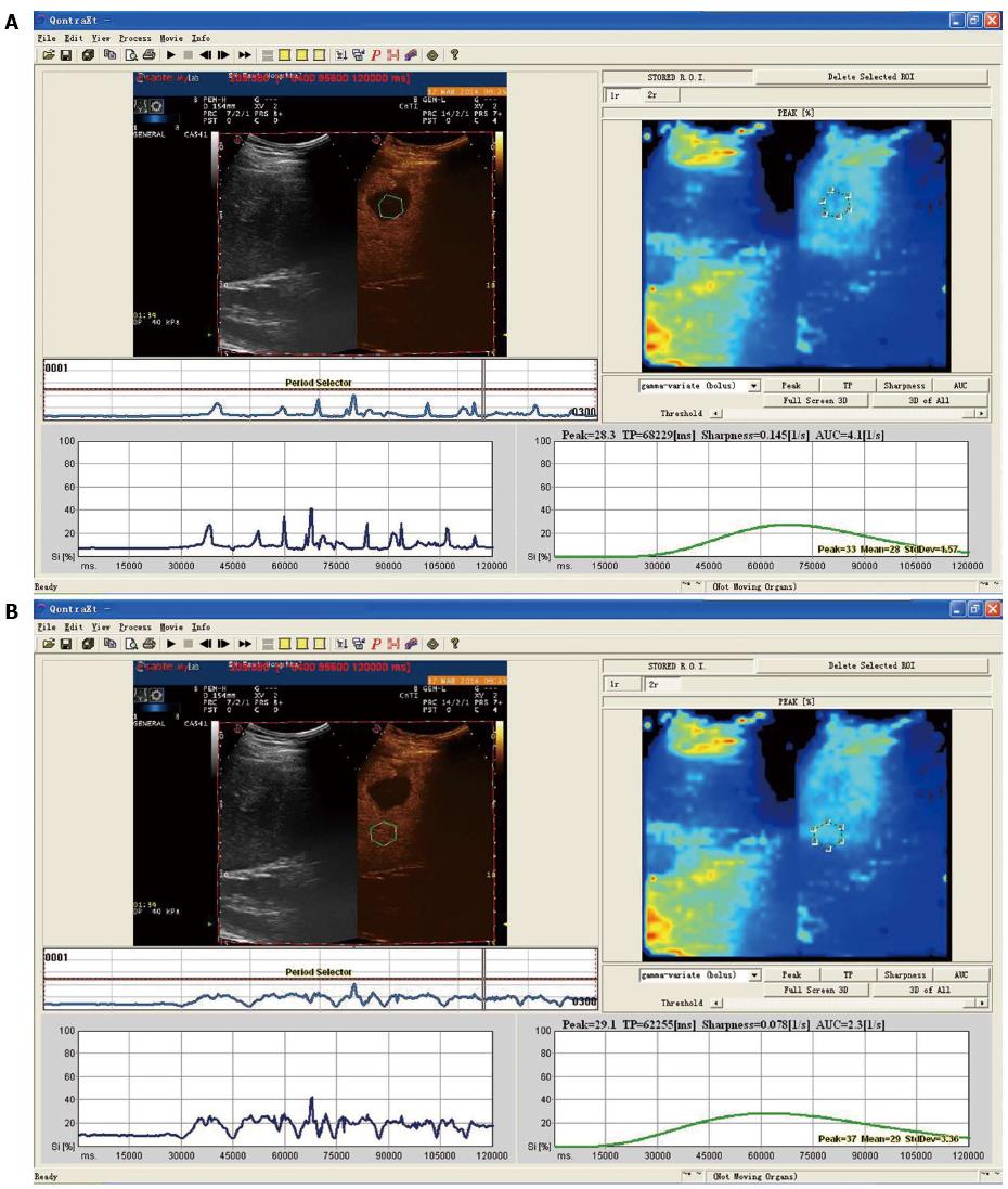
Figure 1 Time-intensity curve of patients in the non-recurrence group.
A: Time-intensity curve of ROI in the tumor. The green curve was gentle, and indicated that the change in intensity was less obvious. The Peak was 28.3% and TP was 68.23 s; B: Time-intensity curve of ROI beyond the tumor. The green curve was gentle and similar to the ROI curve in the tumor. The Peak was 29.1% and TP was 62.26 s. ROI: Region of interest.
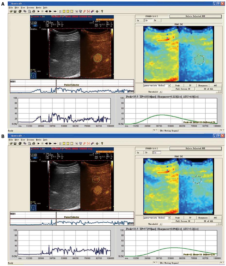
Figure 2 Time-intensity curve of patients in the recurrence group.
A: Time-intensity curve of ROI in the tumor. The green curve was steep, and indicated that the change in intensity was obvious. The Peak was 35.5% and TP was 43.33 s; B: Time-intensity curve of ROI beyond the tumor. The green curve was gentler, compared with the ROI curve in the tumor. The Peak was 33.5% and TP was 57.29 s. ROI: Region of interest.
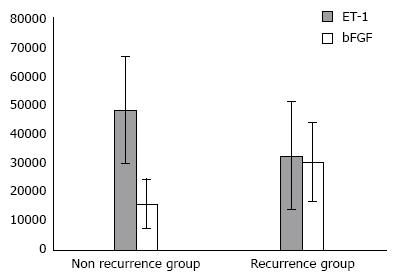
Figure 3 Endothelin-1 and basic fibroblast growth factor expression levels in the non-recurrence and recurrence groups.
ET-1: Endothelin-1; bFGF: Basic fibroblast growth factor.
- Citation: Gao Y, Zheng DY, Cui Z, Ma Y, Liu YZ, Zhang W. Predictive value of quantitative contrast-enhanced ultrasound in hepatocellular carcinoma recurrence after ablation. World J Gastroenterol 2015; 21(36): 10418-10426
- URL: https://www.wjgnet.com/1007-9327/full/v21/i36/10418.htm
- DOI: https://dx.doi.org/10.3748/wjg.v21.i36.10418


