Copyright
©The Author(s) 2015.
World J Gastroenterol. Jul 28, 2015; 21(28): 8569-8579
Published online Jul 28, 2015. doi: 10.3748/wjg.v21.i28.8569
Published online Jul 28, 2015. doi: 10.3748/wjg.v21.i28.8569
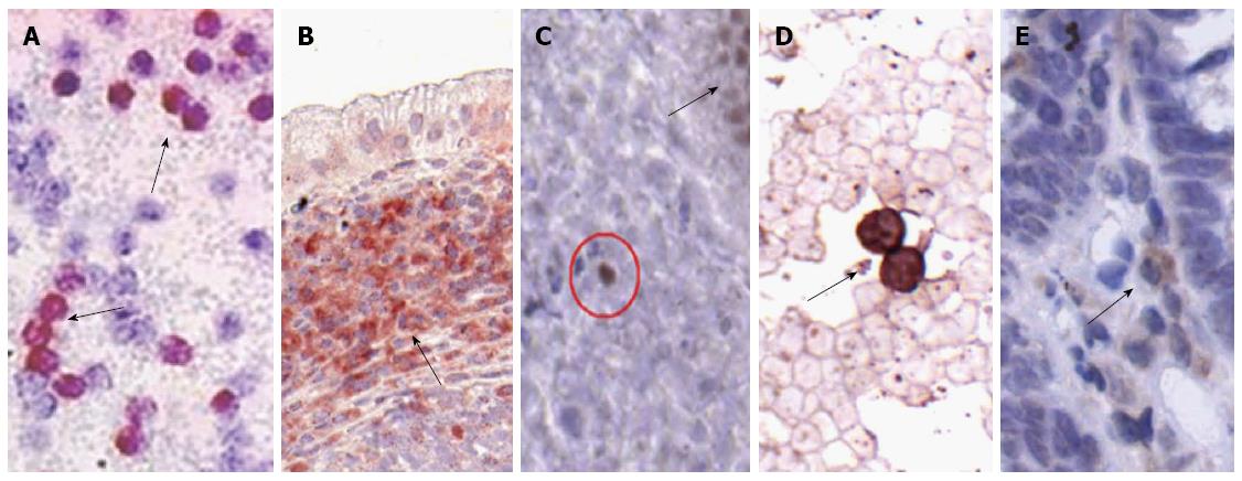
Figure 1 Immunopositive cells in peripheral blood and lamina propria of patients with ulcerative colitis.
A: Hepatocyte-derived growth factor receptor (HGFR)+ cells in peripheral blood (black arrows indicate immunoreactive cells; magnification × 400); B: HGFR+ cells in lamina propria (black arrow indicates a group of immunoreactive cells underneath the epithelium; magnification × 200); C: CDX2+ (clone: ATM28) cell (red circle) in lamina propria (black arrow indicates a crypt base with CDX2+ epithelial cells; magnification × 400); D: CD133+ cells in peripheral blood (black arrow indicates immunoreactive cells; magnification × 600); E: CD133+ cell (black arrow) in lamina propria underneath the epithelium (magnification × 300). In all cases hematoxylin co-staining was performed.
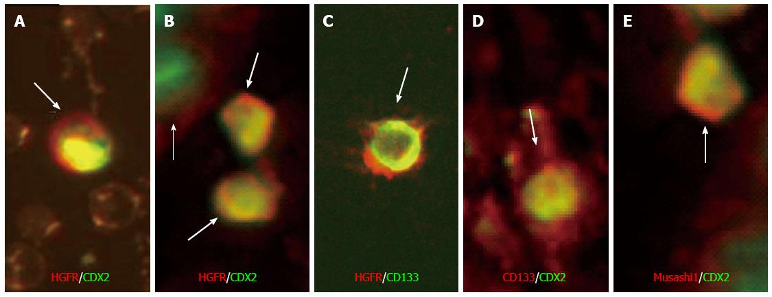
Figure 2 Double fluorescent immunoreactive cells in peripheral blood and lamina propria of patients with active ulcerative colitis.
A: Hepatocyte-derived growth factor receptor (HGFR)+/CDX2+ cell in peripheral blood (white arrow indicates immunoreactive cell; magnification × 600); B: HGFR+/CDX2+ cells in lamina propria (white arrows indicate immunoreactive cells underneath the epithelium; white thin arrow indicates a crypt base with CDX2+ epithelial cell; magnification × 600); C: HGFR+/CD133+ cell in lamina propria (white arrow; magnification × 600); D: CD133+/CDX2+ cell in lamina propria (white arrow; magnification × 600); E: Musashi-1+/CDX2+ cell in the lamina propria (white arrow; magnification × 600). Red fluorescent labeling: Texas-Red; Green fluorescent labeling: FITC.
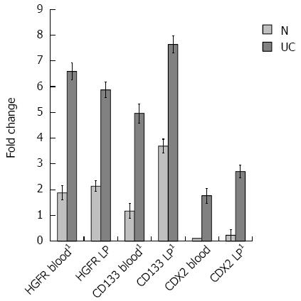
Figure 3 Foldchange alterations of the assayed genes in peripheral blood and lamina propria samples.
1Significant (P < 0.005) gene expression alterations between ulcerative colitis (UC) and healthy control (N) samples. LP: Lamina propria; HGFR: Hepatocyte-derived growth factor receptor.
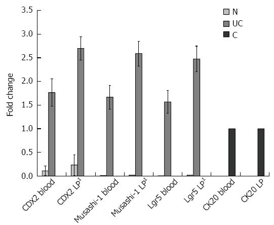
Figure 4 Foldchange alterations of the assayed epithelial genes in peripheral blood and lamina propria samples.
1Significant (P < 0.005) gene expression alterations between ulcerative colitis (UC) and healthy control (N) samples. C: Control samples (SW480 cells in blood, crypt epithelial cells in lamina propria (LP) laser-microdissected samples).
- Citation: Sipos F, Constantinovits M, Valcz G, Tulassay Z, Műzes G. Association of hepatocyte-derived growth factor receptor/caudal type homeobox 2 co-expression with mucosal regeneration in active ulcerative colitis. World J Gastroenterol 2015; 21(28): 8569-8579
- URL: https://www.wjgnet.com/1007-9327/full/v21/i28/8569.htm
- DOI: https://dx.doi.org/10.3748/wjg.v21.i28.8569


