©The Author(s) 2015.
World J Gastroenterol. Jul 21, 2015; 21(27): 8462-8466
Published online Jul 21, 2015. doi: 10.3748/wjg.v21.i27.8462
Published online Jul 21, 2015. doi: 10.3748/wjg.v21.i27.8462
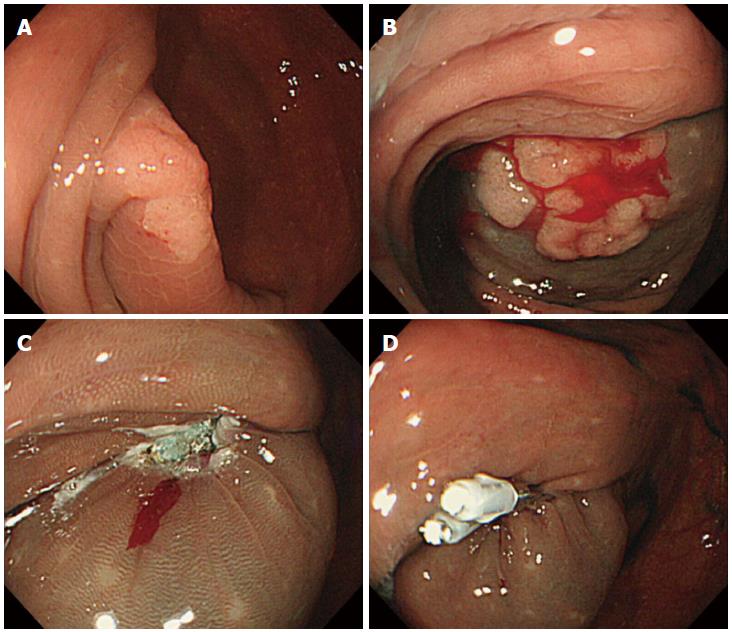
Figure 1 Endoscopic views.
A: A large IIa lesion [laterally spreading tumor (LST) of the granular type, or LST-G lesion located in the cecum. The tumor was estimated to be 15 -mm in diameter; B: After an injection of glycerol into the submucosal layer, the tumor nearly obstructed the orifice of the appendix; C: Ulcer appearance after successful endoscopic mucosal resection, without any complications; D: The ulcer was sutured using two clips, and the appendiceal orifice was avoided.
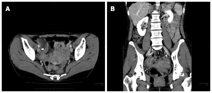
Figure 2 Abdominal contrast-enhanced computed tomography showing appendiceal wall thickening and swelling.
A: Axial section; B: Sagittal section.
- Citation: Nemoto Y, Tokuhisa J, Shimada N, Gomi T, Maetani I. Acute appendicitis following endoscopic mucosal resection of cecal adenoma. World J Gastroenterol 2015; 21(27): 8462-8466
- URL: https://www.wjgnet.com/1007-9327/full/v21/i27/8462.htm
- DOI: https://dx.doi.org/10.3748/wjg.v21.i27.8462


