©The Author(s) 2015.
World J Gastroenterol. Mar 28, 2015; 21(12): 3750-3754
Published online Mar 28, 2015. doi: 10.3748/wjg.v21.i12.3750
Published online Mar 28, 2015. doi: 10.3748/wjg.v21.i12.3750
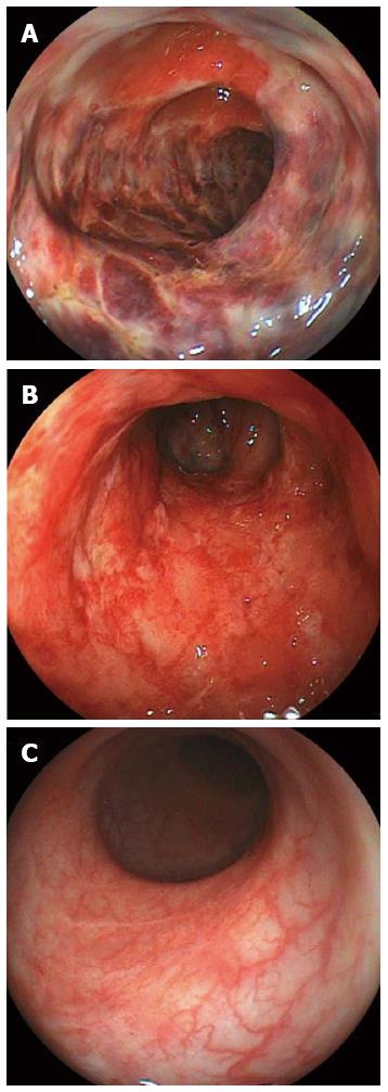
Figure 1 Endoscopic findings.
A: First colonoscopy (day 4), mucosal hyperemic change with edema, erosion and ulcerations and hemorrhagic friable mucosa from the descending to sigmoid colon; B: Follow-up colonoscopy after conservative therapy (day 25); ulcerations remain; C: Follow-up colonoscopy after ganciclovir therapy (day 46); ulcerations have healed.
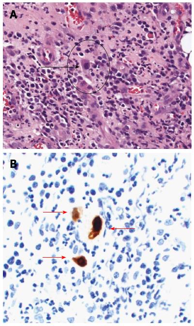
Figure 2 Pathological examinations following hematoxylin-eosin and immunohistochemical staining.
A: White arrow shows cytomegalovirus inclusion bodies (HE staining, × 200); B: Red arrows show cytomegalovirus-positive cells (immunohistochemical staining, × 200).
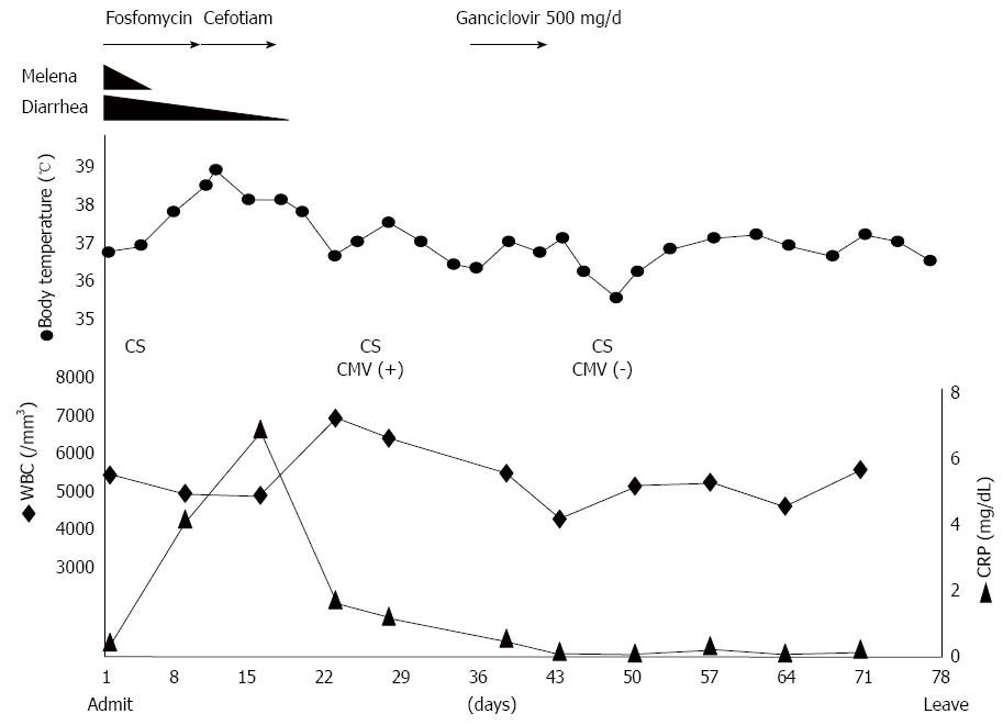
Figure 3 Clinical course of this case.
CS: Colonoscopy; CMV: Cytomegalovirus; CRP: C-reactive protein; WBC: White blood cells.
- Citation: Hasegawa T, Aomatsu K, Nakamura M, Aomatsu N, Aomatsu K. Cytomegalovirus colitis followed by ischemic colitis in a non-immunocompromised adult: A case report. World J Gastroenterol 2015; 21(12): 3750-3754
- URL: https://www.wjgnet.com/1007-9327/full/v21/i12/3750.htm
- DOI: https://dx.doi.org/10.3748/wjg.v21.i12.3750


