©The Author(s) 2015.
World J Gastroenterol. Mar 14, 2015; 21(10): 3127-3131
Published online Mar 14, 2015. doi: 10.3748/wjg.v21.i10.3127
Published online Mar 14, 2015. doi: 10.3748/wjg.v21.i10.3127
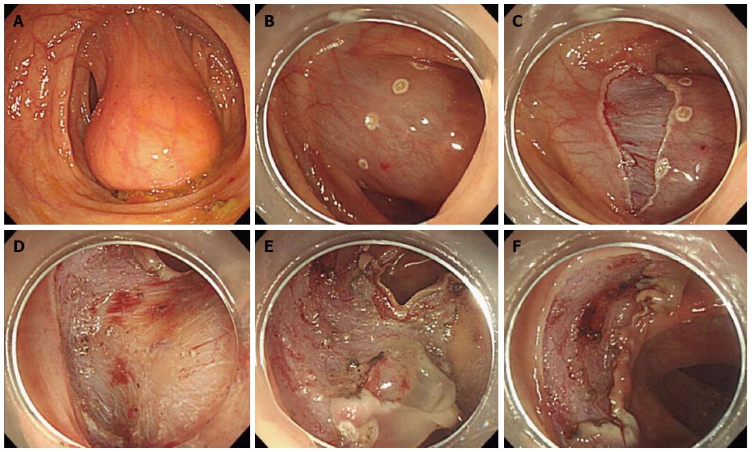
Figure 1 A colonoscopy revealed a soft and yellowish submucosal tumor.
A: Colonoscopic view revealed an approximate 5 cm, yellowish, smooth submucosal tumor on the distal descending colon. B: Markings were made around the tumor on the distal side (anal side) with a dual knife and a lifting solution was injected to make a sufficient submucosal cushion from the muscle layer. C: A precut incision was made on the distal side of the tumor. D: Submucosal dissection was performed with a dual knife, and dissection was done from the distal side (anal side) while visualizing the yellowish tumor surface. E: The tumor was completely dissected from the submucosal layer with the proximal side (oral side) of the mucosal layer remaining. F: The tumor was resected completely without complications.
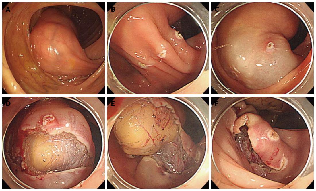
Figure 2 A colonoscopy revealed a yellowish submucosal mass approximately.
A: Colonoscopic view showed an approximate 7 cm, yellowish, smooth submucosal tumor on the proximal ascending colon. B: Markings were made around the tumor on the distal side (anal side) with a dual knife; C: A lifting solution was injected to form a sufficient submucosal cushion. D: Precut incision on the distal side and submucosal dissection were performed with the dual knife. E: Dissection was done with dual and hook knives from the distal side (anal side). F: The tumor was resected as en bloc without complications.
- Citation: Lee JM, Kim JH, Kim M, Kim JH, Lee YB, Lee JH, Lim CW. Endoscopic submucosal dissection of a large colonic lipoma: Report of two cases. World J Gastroenterol 2015; 21(10): 3127-3131
- URL: https://www.wjgnet.com/1007-9327/full/v21/i10/3127.htm
- DOI: https://dx.doi.org/10.3748/wjg.v21.i10.3127


