©The Author(s) 2015.
World J Gastroenterol. Mar 14, 2015; 21(10): 3114-3120
Published online Mar 14, 2015. doi: 10.3748/wjg.v21.i10.3114
Published online Mar 14, 2015. doi: 10.3748/wjg.v21.i10.3114
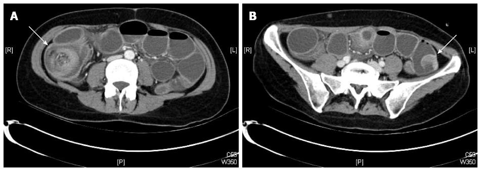
Figure 1 Computed tomography scan findings.
A: Depiction of the ileocolic intussusception (arrow); B: Intraluminal tumor of the small intestine (arrow).
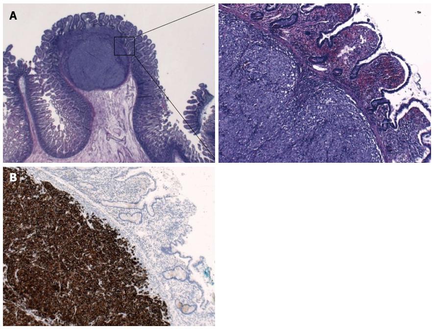
Figure 2 Histopathologic findings.
A: Hematoxylin and eosin stain showing a melanocytic intestinal lesion (left: ×10; right: ×100); B: Immunohistochemical depiction of a melanocytic intestinal lesion with Melan A (×100).
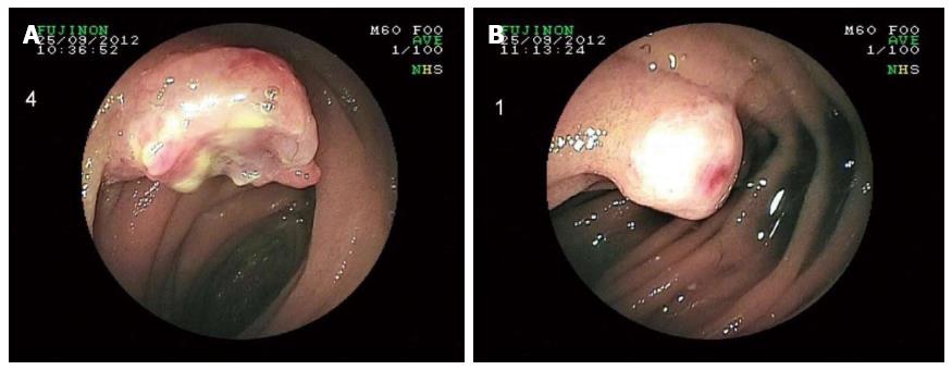
Figure 3 Melanocytic lesions in the proximal jejunum in a double-balloon enteroscopy.
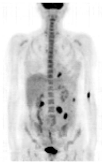
Figure 4 Positron emission tomography-computed tomography scan findings.
Multiple intra-abdominal lesions and a focal fluorodeoxyglucose-accumulating lesion adjacent to the left diaphragm are shown in a maximum intensity projection.
- Citation: Kouladouros K, Gärtner D, Münch S, Paul M, Schön MR. Recurrent intussusception as initial manifestation of primary intestinal melanoma: Case report and literature review. World J Gastroenterol 2015; 21(10): 3114-3120
- URL: https://www.wjgnet.com/1007-9327/full/v21/i10/3114.htm
- DOI: https://dx.doi.org/10.3748/wjg.v21.i10.3114


